Department of Ophthalmology, Faculty of Medicine and University Hospital Cologne, University of Cologne, Kerpener Strasse 62, 50937, Cologne, Germany.
Eye Center, Second Affiliated Hospital, School of Medicine, Zhejiang University, Hangzhou, Zhejiang, China.
Aesthetic Plast Surg. 2021 Aug;45(4):1601-1610. doi: 10.1007/s00266-020-02091-5. Epub 2021 Jan 15.
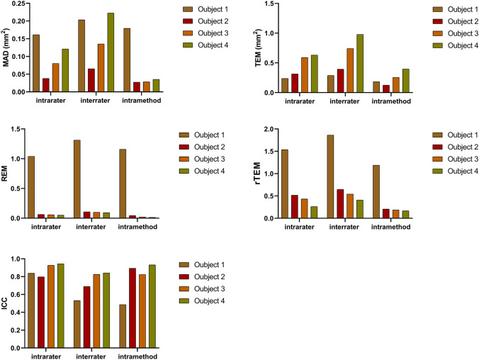
Three-dimensional (3D) stereophotography area measurements are essential for describing morphology in the periocular region. However, its reliability has not yet been sufficiently validated. The objective of this study was to evaluate the reliability of 3D stereophotogrammetric area measurements in the periocular region. Forty healthy volunteers had five flat paper objects placed at each of the seven periocular positions including the endocanthion and the upper medial, upper middle, upper lateral, lower medial, lower middle, and the lower lateral eyelid. Two series of photographic images were captured twice by the same investigator. Each image of the first series was measured twice by the same rater, while images of both series were measured once by a second rater. Differences between these measurements were calculated, and the intrarater, interrater, and intramethod reliability was evaluated for intraclass correlation coefficients (ICCs), mean absolute differences (MADs), technical errors of measurements (TEMs), relative errors of measurements (REMs), and relative TEM (rTEM). Our results showed that 21.2% of all ICCs were considered as excellent, 45.5% were good, 27.3% were moderate, and 6.1% were poor. The interrater ICC for the endocanthion location was 0.4% on a low level. MAD values for all objects were less than 0.3 mm, all TEM were less than 1 mm, the REM and rTEM were less than 2% for all objects, showing high reliability. 3D stereophotogrammetry is a highly reliable system for periocular area measurements and may be used in the clinical routine for planning oculoplastic surgeries and for evaluating changes in periocular morphology.Level of Evidence IV This journal requires that authors assign a level of evidence to each article. For a full description of these Evidence-Based Medicine ratings, please refer to the Table of Contents or the online Instructions to Authors www.springer.com/00266.
三维(3D)立体摄影面积测量对于描述眼眶区域的形态至关重要。然而,其可靠性尚未得到充分验证。本研究旨在评估 3D 立体摄影在眼眶区域面积测量中的可靠性。
四十名健康志愿者的眼眶周围七个位置(包括内眦和上内侧、上中、上外侧、下内侧、下中、下外侧)各放置五个平面纸物体。由同一位研究者拍摄两次系列的照片。第一次系列的每张照片均由同一位评估者测量两次,而两次系列的照片则由第二位评估者测量一次。计算这些测量值之间的差异,并评估组内、组间和组内测量的内在信度,采用组内相关系数(ICC)、平均绝对差值(MAD)、测量技术误差(TEM)、测量相对误差(REM)和相对 TEM(rTEM)进行评估。
我们的结果显示,所有 ICC 中有 21.2%被认为是优秀的,45.5%是良好的,27.3%是中等的,6.1%是较差的。内眦位置的组间 ICC 处于低水平,为 0.4%。所有物体的 MAD 值均小于 0.3 毫米,所有 TEM 值均小于 1 毫米,所有物体的 REM 和 rTEM 值均小于 2%,显示出高度的可靠性。
3D 立体摄影是一种非常可靠的眼眶区域测量系统,可用于眼整形手术的临床常规规划,并用于评估眼眶形态的变化。
证据等级 IV 本杂志要求作者为每篇文章分配一个证据等级。有关这些循证医学评级的完整描述,请参阅目录或在线作者指南 www.springer.com/00266。