• 文献检索
  • 文档翻译
  • 深度研究
  • 学术资讯
  • Suppr Zotero 插件Zotero 插件
  • 邀请有礼
  • 套餐&价格
  • 历史记录
应用&插件
Suppr Zotero 插件Zotero 插件浏览器插件Mac 客户端Windows 客户端微信小程序
定价
高级版会员购买积分包购买API积分包
服务
文献检索文档翻译深度研究API 文档MCP 服务
关于我们
关于 Suppr公司介绍联系我们用户协议隐私条款
关注我们

Suppr 超能文献

核心技术专利:CN118964589B侵权必究
粤ICP备2023148730 号-1Suppr @ 2026

文献检索

告别复杂PubMed语法,用中文像聊天一样搜索,搜遍4000万医学文献。AI智能推荐,让科研检索更轻松。

立即免费搜索

文件翻译

保留排版,准确专业,支持PDF/Word/PPT等文件格式,支持 12+语言互译。

免费翻译文档

深度研究

AI帮你快速写综述,25分钟生成高质量综述,智能提取关键信息,辅助科研写作。

立即免费体验

三维磁共振成像(3D MRI)在诊断距腓前韧带损伤中的应用评估。

Evaluation of 3-Dimensional Magnetic Resonance Imaging (3D MRI) in Diagnosing Anterior Talofibular Ligament Injury.

机构信息

Department of Orthopedics, Union Hospital, Tongji Medical College, Huazhong University of Science and Technology, Wuhan, Hubei, China (mainland).

Department of Orthopedic Surgery, Shanghai Key Laboratory of Orthopedic Implants, Shanghai Ninth People's Hospital, Shanghai Jiaotong University School of Medicine, Shanghai, China (mainland).

出版信息

Med Sci Monit. 2021 Jan 16;27:e927920. doi: 10.12659/MSM.927920.

DOI:10.12659/MSM.927920
PMID:33453097
原文链接:https://pmc.ncbi.nlm.nih.gov/articles/PMC7816539/
Abstract

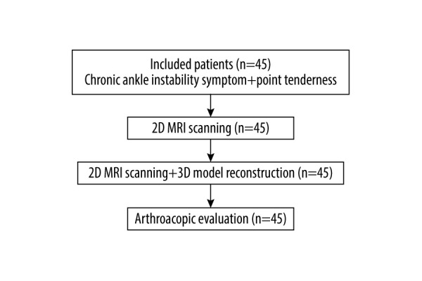
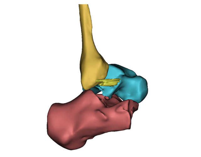
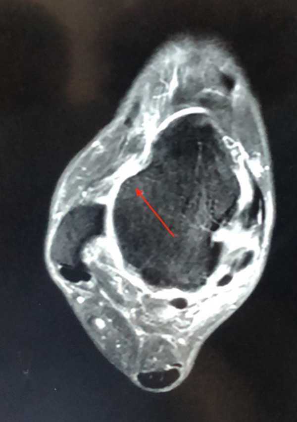
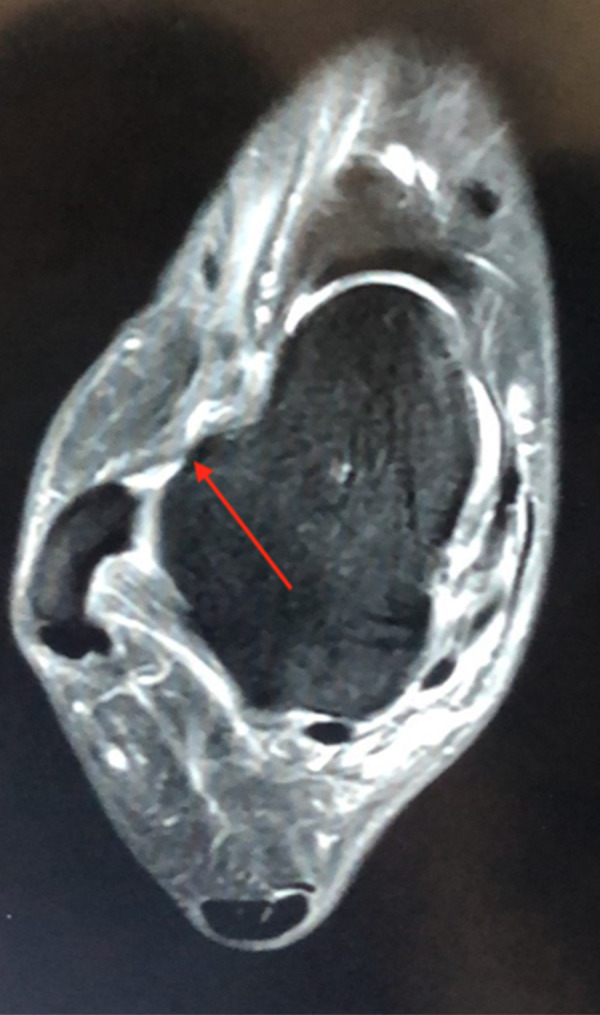
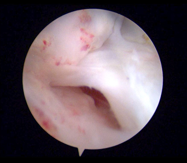
BACKGROUND It is challenging to entirely show the anterior talofibular ligament (ATFL) and accurately diagnose ATFL injury with traditional 2-dimensional (2D) magnetic resonance imaging (MRI). With the introduction of 3.0T MRI, a 3-dimensional (3D) MRI sequence can achieve images with high spatial resolution. This study aimed to evaluate the accuracy of 3D MRI and compare it with 2D MRI in diagnosing ATFL injury. MATERIAL AND METHODS This was a prospective study in which 45 patients with clinically suspected ATFL injury underwent 2D MRI, 3D MRI, and 3D model reconstruction followed by arthroscopic surgery between February 2018 and April 2019. Two radiologists who had over 11 and 13 years of musculoskeletal experience assessed the injury of ATFL in consensus without any clinical clues. Arthroscopic surgery results were the standard reference of MRI accuracy. RESULTS The 3D MRI results of ATFL injury showed the sensitivity of diagnosis of complete tears of 83% and specificity of 82%. The partial tears diagnosis sensitivity was 78%, and specificity was 100%. The sensitivity of diagnosis of sprains was 100%, and the specificity was 97%. The 3D MRI accuracy of diagnosis was 98% for no injury, 98% for sprain, 91% for partial tear, and 82% for complete tear. The difference in the diagnosis of sprain and partial tears by 3D MRI and 2D MRI was statistically significant (P<0.05). A 3D reconstruction model was successfully created for all patients. CONCLUSIONS 3D MRI may be a reliable and accurate method to detect ATFL injury. The 3D reconstruction model using 3D MRI sequences has excellent prospects in application.

摘要

背景

传统二维(2D)磁共振成像(MRI)在完全显示距腓前韧带(ATFL)和准确诊断 ATFL 损伤方面具有挑战性。随着 3.0T MRI 的引入,三维(3D)MRI 序列可以实现高空间分辨率的图像。本研究旨在评估 3D MRI 的准确性,并将其与 2D MRI 诊断 ATFL 损伤进行比较。

材料和方法

这是一项前瞻性研究,2018 年 2 月至 2019 年 4 月期间,45 例临床疑似 ATFL 损伤的患者接受了 2D MRI、3D MRI 和 3D 模型重建,随后进行了关节镜手术。两名具有超过 11 年和 13 年肌肉骨骼经验的放射科医生在没有任何临床线索的情况下进行了共识评估。关节镜手术结果是 MRI 准确性的标准参考。

结果

3D MRI 对 ATFL 损伤的诊断结果显示,完全撕裂的诊断敏感性为 83%,特异性为 82%。部分撕裂的诊断敏感性为 78%,特异性为 100%。扭伤的诊断敏感性为 100%,特异性为 97%。无损伤、扭伤、部分撕裂和完全撕裂的 3D MRI 诊断准确率分别为 98%、98%、91%和 82%。3D MRI 和 2D MRI 诊断扭伤和部分撕裂的差异有统计学意义(P<0.05)。所有患者均成功创建了 3D 重建模型。

结论

3D MRI 可能是一种可靠且准确的检测 ATFL 损伤的方法。使用 3D MRI 序列的 3D 重建模型在应用方面具有广阔的前景。

https://cdn.ncbi.nlm.nih.gov/pmc/blobs/ebc6/7816539/9d5c45dd0100/medscimonit-27-e927920-g005.jpg
https://cdn.ncbi.nlm.nih.gov/pmc/blobs/ebc6/7816539/834015a8ead0/medscimonit-27-e927920-g001.jpg
https://cdn.ncbi.nlm.nih.gov/pmc/blobs/ebc6/7816539/8927ba9513bf/medscimonit-27-e927920-g002.jpg
https://cdn.ncbi.nlm.nih.gov/pmc/blobs/ebc6/7816539/001fb4161f59/medscimonit-27-e927920-g003.jpg
https://cdn.ncbi.nlm.nih.gov/pmc/blobs/ebc6/7816539/2db17965c967/medscimonit-27-e927920-g004.jpg
https://cdn.ncbi.nlm.nih.gov/pmc/blobs/ebc6/7816539/9d5c45dd0100/medscimonit-27-e927920-g005.jpg

相似文献

1
Evaluation of 3-Dimensional Magnetic Resonance Imaging (3D MRI) in Diagnosing Anterior Talofibular Ligament Injury.三维磁共振成像(3D MRI)在诊断距腓前韧带损伤中的应用评估。
Med Sci Monit. 2021 Jan 16;27:e927920. doi: 10.12659/MSM.927920.
2
Three-dimensional isotropic T2-weighted fast spin-echo (VISTA) ankle MRI versus two-dimensional fast spin-echo T2-weighted sequences for the evaluation of anterior talofibular ligament injury.三维各向同性T2加权快速自旋回波(VISTA)踝关节磁共振成像与二维快速自旋回波T2加权序列在评估距腓前韧带损伤中的比较
Clin Radiol. 2016 Apr;71(4):349-55. doi: 10.1016/j.crad.2015.12.007. Epub 2016 Jan 14.
3
Accuracy of MRI findings in chronic lateral ankle ligament injury: comparison with surgical findings.MRI 检查在慢性外侧踝关节韧带损伤中的准确性:与手术结果的比较。
Clin Radiol. 2012 Apr;67(4):313-8. doi: 10.1016/j.crad.2011.08.025. Epub 2011 Nov 9.
4
Evaluation of the anterior inferior tibiofibular and anterior talofibular ligaments using 2D oblique coronal imaging and 3D isotropic resolution T2-weighted fast spin-echo sequences at 3.0 T: Is there additional diagnostic value?在 3.0T 磁共振中使用二维斜冠状成像和三维各向同性分辨率 T2 加权快速自旋回波序列评估下胫腓前韧带和前距腓韧带:是否有额外的诊断价值?
J Med Imaging Radiat Oncol. 2020 Jun;64(3):338-346. doi: 10.1111/1754-9485.13028. Epub 2020 Apr 1.
5
MRI of the anterior talofibular ligament, talar cartilage and os subfibulare: Comparison of isotropic resolution 3D and conventional 2D T2-weighted fast spin-echo sequences at 3.0 T.距腓前韧带、距骨软骨和腓骨下骨的磁共振成像:3.0T下各向同性分辨率三维与传统二维T2加权快速自旋回波序列的比较
Skeletal Radiol. 2016 Jul;45(7):899-908. doi: 10.1007/s00256-016-2367-x. Epub 2016 Mar 19.
6
[Lesions of the collateral ligaments of the ankle: diagnosis and follow-up with magnetic resonance and ultrasonography].[踝关节侧副韧带损伤:磁共振成像与超声检查的诊断及随访]
Radiol Med. 1996 Jun;91(6):705-9.
7
Reliability and Validity of Magnetic Resonance Imaging for the Evaluation of the Anterior Talofibular Ligament in Patients Undergoing Ankle Arthroscopy.踝关节镜检查患者中磁共振成像评估距腓前韧带的可靠性和有效性
Arthroscopy. 2015 Aug;31(8):1540-7. doi: 10.1016/j.arthro.2015.02.024. Epub 2015 Apr 14.
8
3D MRI evaluation of morphological characteristics of lateral ankle ligaments in injured patients and uninjured controls.3D MRI对受伤患者和未受伤对照者外侧踝关节韧带形态特征的评估。
J Orthop Sci. 2020 Jan;25(1):183-187. doi: 10.1016/j.jos.2019.02.018. Epub 2019 Mar 16.
9
Increased ATFL-PTFL angle could be an indirect MRI sign in diagnosis of chronic ATFL injury.距腓前韧带-跟腓韧带夹角增大可能是慢性距腓前韧带损伤的间接 MRI 征象。
Knee Surg Sports Traumatol Arthrosc. 2020 Jan;28(1):208-212. doi: 10.1007/s00167-018-5252-4. Epub 2018 Nov 9.
10
The anterior talofibular ligament: A thin-slice three-dimensional magnetic resonance imaging study.距腓前韧带:一项薄层三维磁共振成像研究。
Foot Ankle Surg. 2022 Dec;28(8):1202-1209. doi: 10.1016/j.fas.2021.11.011. Epub 2021 Dec 2.

引用本文的文献

1
Development and validation of radiomics model for MRI-based identification of anterior talofibular ligament injuries.基于MRI的距腓前韧带损伤识别的影像组学模型的开发与验证
Sci Rep. 2025 May 4;15(1):15575. doi: 10.1038/s41598-025-99813-z.
2
Chronic ankle instability: a cadaveric anatomical and 3D high-resolution MRI study for surgical reconstruction procedures.慢性踝关节不稳:一项用于手术重建程序的尸体解剖和三维高分辨率磁共振成像研究
Insights Imaging. 2024 Oct 14;15(1):249. doi: 10.1186/s13244-024-01824-3.
3
Ultrasound or MRI in the Evaluation of Anterior Talofibular Ligament (ATFL) Injuries: Systematic Review and Meta-Analysis.

本文引用的文献

1
3D MRI evaluation of morphological characteristics of lateral ankle ligaments in injured patients and uninjured controls.3D MRI对受伤患者和未受伤对照者外侧踝关节韧带形态特征的评估。
J Orthop Sci. 2020 Jan;25(1):183-187. doi: 10.1016/j.jos.2019.02.018. Epub 2019 Mar 16.
2
Reliability and validity of different ankle MRI scanning planes for the anterior talofibular ligament injury diagnosis: a cadaveric study.不同踝关节MRI扫描平面在前距腓韧带损伤诊断中的可靠性和有效性:一项尸体研究
J Orthop Surg Res. 2019 Feb 28;14(1):69. doi: 10.1186/s13018-019-1102-4.
3
3D Visualization and Augmented Reality for Orthopedics.
超声或磁共振成像在评估距腓前韧带(ATFL)损伤中的应用:系统评价与荟萃分析
Diagnostics (Basel). 2023 Jul 10;13(14):2324. doi: 10.3390/diagnostics13142324.
4
The increased anterior talofibular ligament-posterior talofibular ligament angle on MRI may help evaluate chronic ankle instability.MRI 上增加的距腓前韧带-距腓后韧带夹角有助于评估慢性踝关节不稳定。
Surg Radiol Anat. 2023 Oct;45(10):1205-1211. doi: 10.1007/s00276-023-03196-7. Epub 2023 Jul 10.
5
Role of the intrinsic subtalar ligaments in subtalar instability and consequences for clinical practice.距下关节内在韧带在距下关节不稳定中的作用及对临床实践的影响。
Front Bioeng Biotechnol. 2023 Mar 10;11:1047134. doi: 10.3389/fbioe.2023.1047134. eCollection 2023.
3D 可视化与增强现实技术在骨科中的应用。
Adv Exp Med Biol. 2018;1093:193-205. doi: 10.1007/978-981-13-1396-7_16.
4
3D printing- creating a blueprint for the future of orthopedics: Current concept review and the road ahead!3D打印——为骨科未来绘制蓝图:当前概念综述及未来之路!
J Clin Orthop Trauma. 2018 Jul-Sep;9(3):207-212. doi: 10.1016/j.jcot.2018.07.007. Epub 2018 Jul 23.
5
Accuracy of magnetic resonance imaging in diagnosing lateral ankle ligament injuries: A comparative study with surgical findings and timings of scans.磁共振成像诊断外侧踝关节韧带损伤的准确性:与手术结果及扫描时机的对比研究
Asia Pac J Sports Med Arthrosc Rehabil Technol. 2016 Nov 20;7:15-20. doi: 10.1016/j.asmart.2016.09.003. eCollection 2017 Jan.
6
Subtalar instability: imaging features of subtalar ligaments on 3D isotropic ankle MRI.距下关节不稳:3D各向同性踝关节MRI上距下韧带的影像学特征
BMC Musculoskelet Disord. 2017 Nov 21;18(1):475. doi: 10.1186/s12891-017-1841-5.
7
3D printing from MRI Data: Harnessing strengths and minimizing weaknesses.基于磁共振成像数据的3D打印:发挥优势,减少劣势。
J Magn Reson Imaging. 2017 Mar;45(3):635-645. doi: 10.1002/jmri.25526. Epub 2016 Nov 22.
8
MRI of the anterior talofibular ligament, talar cartilage and os subfibulare: Comparison of isotropic resolution 3D and conventional 2D T2-weighted fast spin-echo sequences at 3.0 T.距腓前韧带、距骨软骨和腓骨下骨的磁共振成像:3.0T下各向同性分辨率三维与传统二维T2加权快速自旋回波序列的比较
Skeletal Radiol. 2016 Jul;45(7):899-908. doi: 10.1007/s00256-016-2367-x. Epub 2016 Mar 19.
9
3D Printout Models vs. 3D-Rendered Images: Which Is Better for Preoperative Planning?3D打印模型与3D渲染图像:哪种更适合术前规划?
J Surg Educ. 2016 May-Jun;73(3):518-23. doi: 10.1016/j.jsurg.2016.01.003. Epub 2016 Feb 6.
10
The anterior talofibular ligament: A detailed morphological study.距腓前韧带:一项详细的形态学研究。
Foot (Edinb). 2015 Sep;25(3):141-7. doi: 10.1016/j.foot.2015.05.004. Epub 2015 May 27.