Howard Hughes Medical Institute, Department of Oncological Sciences and Huntsman Cancer Institute, University of Utah School of Medicine, Salt Lake City, UT 84112, USA; Division of Urology, Department of Surgery, University of Utah School of Medicine, Salt Lake City, UT 84112, USA.
Department of Molecular, Cell, and Developmental Biology, University of California, Los Angeles, Los Angeles, CA 90095, USA.
Cell Stem Cell. 2021 Apr 1;28(4):764-778.e4. doi: 10.1016/j.stem.2020.12.004. Epub 2021 Jan 15.
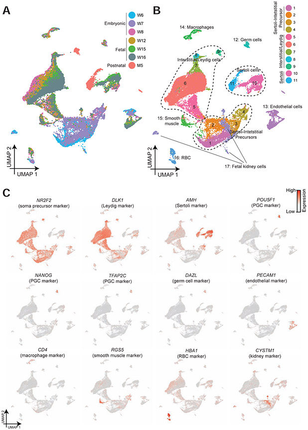
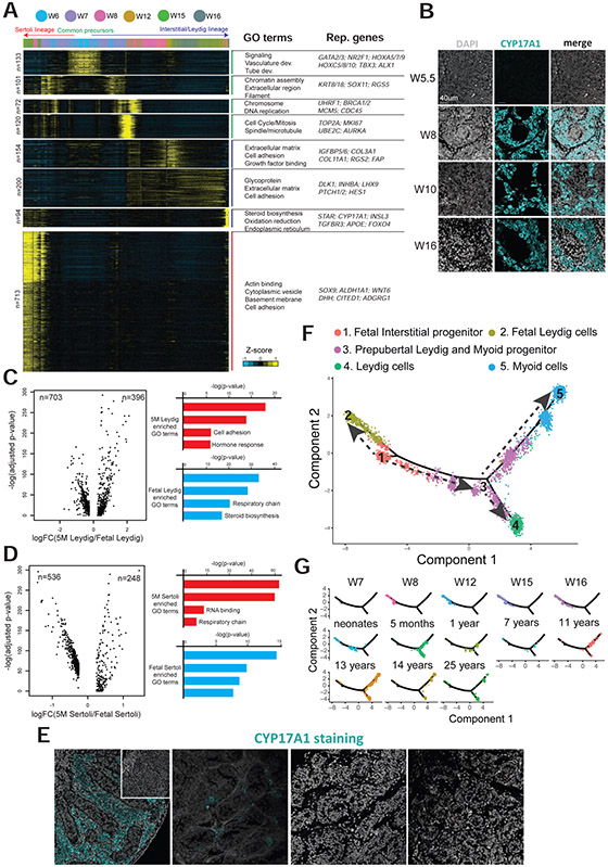
Human testis development in prenatal life involves complex changes in germline and somatic cell identity. To better understand, we profiled and analyzed ∼32,500 single-cell transcriptomes of testicular cells from embryonic, fetal, and infant stages. Our data show that at 6-7 weeks postfertilization, as the testicular cords are established, the Sertoli and interstitial cells originate from a common heterogeneous progenitor pool, which then resolves into fetal Sertoli cells (expressing tube-forming genes) or interstitial cells (including Leydig-lineage cells expressing steroidogenesis genes). Almost 10 weeks later, beginning at 14-16 weeks postfertilization, the male primordial germ cells exit mitosis, downregulate pluripotent transcription factors, and transition into cells that strongly resemble the state 0 spermatogonia originally defined in the infant and adult testes. Therefore, we called these fetal spermatogonia "state f0." Overall, we reveal multiple insights into the coordinated and temporal development of the embryonic, fetal, and postnatal male germline together with the somatic niche.
人类胚胎期睾丸发育涉及生殖细胞和体细胞身份的复杂变化。为了更好地理解这一过程,我们对胚胎期、胎儿期和婴儿期睾丸细胞的约 32500 个单细胞转录组进行了分析。我们的数据表明,在受精后 6-7 周,当睾丸索建立时,支持细胞和间质细胞起源于一个共同的异质祖细胞池,然后分化为胎儿支持细胞(表达形成小管的基因)或间质细胞(包括表达类固醇生成基因的莱迪希谱系细胞)。大约 10 周后,从受精后 14-16 周开始,雄性原始生殖细胞退出有丝分裂,下调多能转录因子,并转化为与婴儿和成年睾丸中最初定义的状态 0 精原细胞非常相似的细胞。因此,我们将这些胎儿精原细胞称为“状态 f0”。总的来说,我们揭示了胚胎期、胎儿期和出生后雄性生殖细胞与体细胞龛之间协调和时间上的发育的多个见解。