Dölen Yusuf, Valente Michael, Tagit Oya, Jäger Eliezer, Van Dinther Eric A W, van Riessen N Koen, Hruby Martin, Gileadi Uzi, Cerundolo Vincenzo, Figdor Carl G
Department of Tumor Immunology, Radboud Institute for Molecular Life Sciences, Radboud University Medical Center & Oncode Institute, Nijmegen, The Netherlands.
Institute of Macromolecular Chemistry V.v.i., Academy of Sciences of the Czech Republic, Prague 6, Czech Republic.
Oncoimmunology. 2020 Mar 17;9(1):1738813. doi: 10.1080/2162402X.2020.1738813.
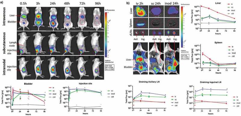
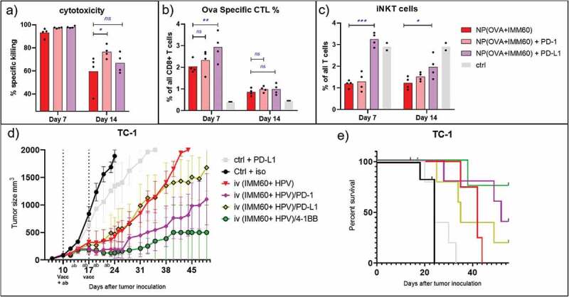
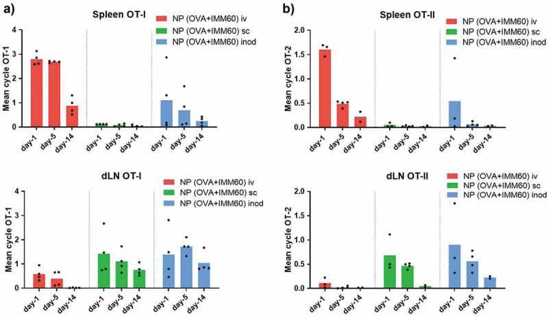
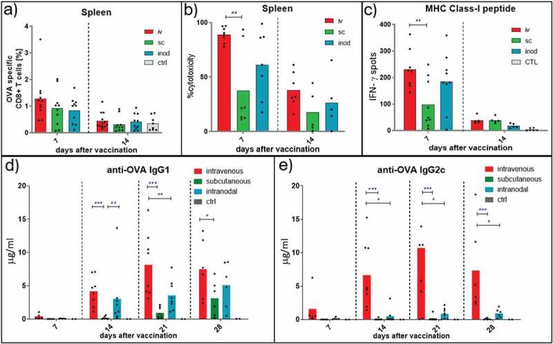
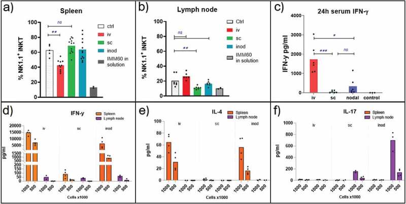
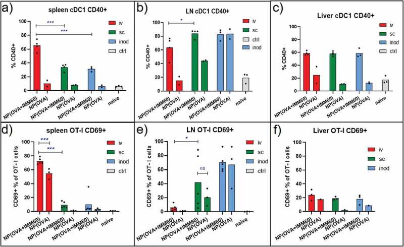
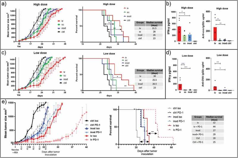
Nanovaccines, co-delivering antigen and invariant natural killer T (iNKT) cell agonists, proved to be very effective in inducing anti-tumor T cell responses due to their exceptional helper function. However, it is known that iNKT cells are not equally present in all lymphoid organs and nanoparticles do not get evenly distributed to all immune compartments. In this study, we evaluated the effect of the vaccination route on iNKT cell help to T and B cell responses for the first time in an antigen and agonist co-delivery setting. Intravenous administration of PLGA nanoparticles was mainly targeting liver and spleen where iNKT1 cells are abundant and induced the highest serum IFN-y levels, T cell cytotoxicity, and Th-1 type antibody responses. In comparison, after subcutaneous or intranodal injections, nanoparticles mostly drained or remained in regional lymph nodes where iNKT17 cells were abundant. After subcutaneous and intranodal injections, antigen-specific IgG2 c production was hampered and IFN-y production, as well as cytotoxic T cell responses, depended on sporadic systemic drainage. Therapeutic anti-tumor experiments also demonstrated a clear advantage of intravenous injection over intranodal or subcutaneous vaccinations. Moreover, tumor control could be further improved by PD-1 immune checkpoint blockade after intravenous vaccination, but not by intranodal vaccination. Anti PD-1 antibody combination mainly exerts its effect by prolonging the cytotoxicity of T cells. Nanovaccines also demonstrated synergism with anti-4-1BB agonistic antibody treatment in controlling tumor growth. We conclude that nanovaccines containing iNKT cell agonists shall be preferentially administered intravenously, to optimally reach cellular partners for inducing effective anti-tumor immune responses.
纳米疫苗可共同递送抗原和不变自然杀伤T(iNKT)细胞激动剂,由于其特殊的辅助功能,在诱导抗肿瘤T细胞反应方面被证明非常有效。然而,已知iNKT细胞并非在所有淋巴器官中均等存在,且纳米颗粒也不会均匀分布到所有免疫区室。在本研究中,我们首次在抗原和激动剂共同递送的情况下,评估了接种途径对iNKT细胞辅助T细胞和B细胞反应的影响。静脉注射聚乳酸-羟基乙酸共聚物(PLGA)纳米颗粒主要靶向肝脏和脾脏,这些器官中iNKT1细胞丰富,并诱导了最高的血清干扰素-γ水平、T细胞细胞毒性和Th-1型抗体反应。相比之下,皮下或淋巴结内注射后,纳米颗粒大多引流至或留存于iNKT17细胞丰富的局部淋巴结中。皮下和淋巴结内注射后,抗原特异性IgG2c的产生受到阻碍,干扰素-γ的产生以及细胞毒性T细胞反应取决于偶发的全身引流。治疗性抗肿瘤实验也证明了静脉注射相对于淋巴结内或皮下接种具有明显优势。此外,静脉接种后通过程序性死亡受体1(PD-1)免疫检查点阻断可进一步改善肿瘤控制,但淋巴结内接种则不然。抗PD-1抗体组合主要通过延长T细胞的细胞毒性发挥作用。纳米疫苗在控制肿瘤生长方面也显示出与抗4-1BB激动剂抗体治疗具有协同作用。我们得出结论,含有iNKT细胞激动剂的纳米疫苗应优先通过静脉注射给药,以最佳方式接触细胞伙伴,从而诱导有效的抗肿瘤免疫反应。