Department of Neurobiology, School of Basic Medical Sciences, Beijing Key Laboratory of Neural Regeneration and Repair, Beijing Institute for Brain Disorders, Capital Medical University, Beijing, 100069, China.
Department of Neurology, Xuanwu Hospital, Capital Medical University, Beijing, 100053, China.
J Neuroinflammation. 2021 Jan 18;18(1):23. doi: 10.1186/s12974-021-02079-1.
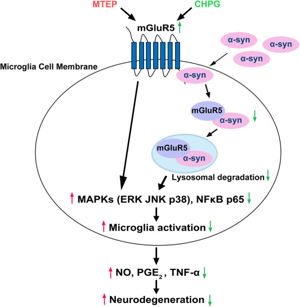
Microglia activation induced by α-synuclein (α-syn) is one of the most important factors in Parkinson's disease (PD) pathogenesis. However, the molecular mechanisms by which α-syn exerts neuroinflammation and neurotoxicity remain largely elusive. Targeting metabotropic glutamate receptor 5 (mGluR5) has been an attractive strategy to mediate microglia activation for neuroprotection, which might be an essential regulator to modulate α-syn-induced neuroinflammation for the treatment of PD. Here, we showed that mGluR5 inhibited α-syn-induced microglia inflammation to protect from neurotoxicity in vitro and in vivo.
Co-immunoprecipitation assays were utilized to detect the interaction between mGluR5 and α-syn in microglia. Griess, ELISA, real-time PCR, western blotting, and immunofluorescence assays were used to detect the regulation of α-syn-induced inflammatory signaling, cytokine secretion, and lysosome-dependent degradation.
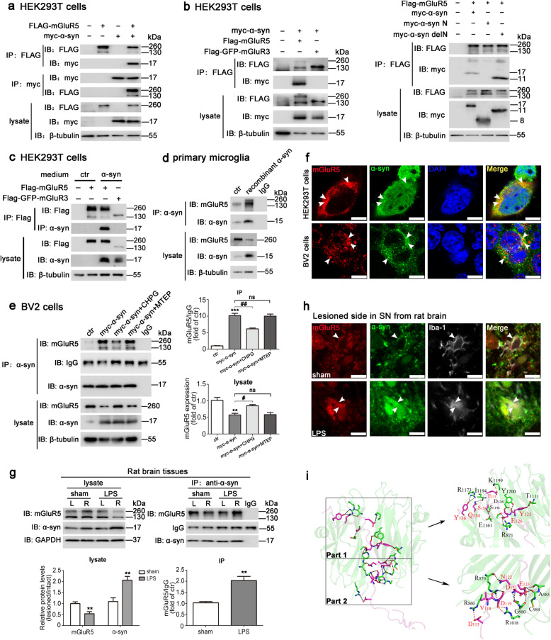
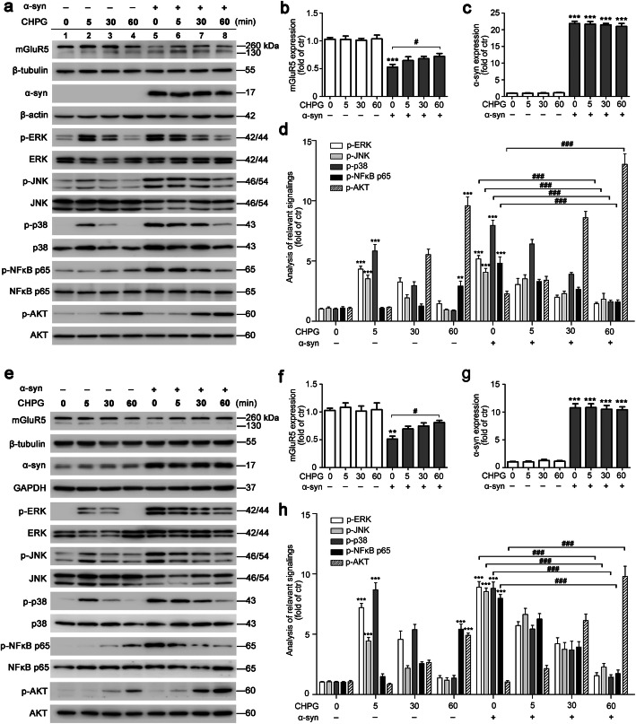
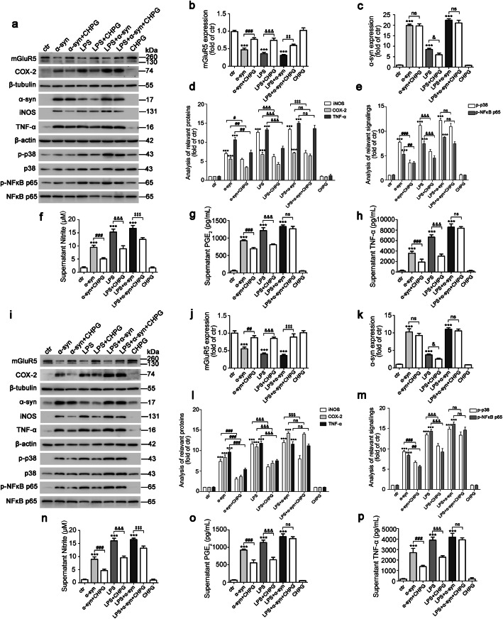
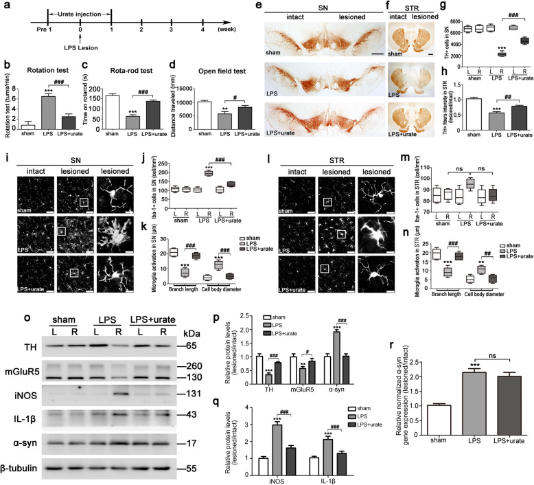
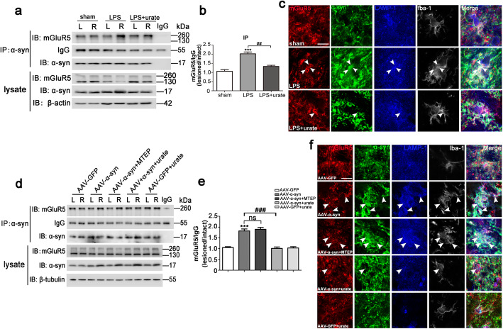
α-syn selectively interacted with mGluR5 but not mGluR3, and α-syn N terminal deletion region was essential for binding to mGluR5 in co-transfected HEK293T cells. The interaction between these two proteins was further detected in BV2 microglia, which was inhibited by the mGluR5 specific agonist CHPG without effect by its selective antagonist MTEP. Moreover, in both BV2 cells and primary microglia, activation of mGluR5 by CHPG partially inhibited α-syn-induced inflammatory signaling and cytokine secretion and also inhibited the microglia activation to protect from neurotoxicity. We further found that α-syn overexpression decreased mGluR5 expression via a lysosomal pathway, as evidenced by the lysosomal inhibitor, NHCl, by blocking mGluR5 degradation, which was not evident with the proteasome inhibitor, MG132. Additionally, co-localization of mGluR5 with α-syn was detected in lysosomes as merging with its marker, LAMP-1. Consistently, in vivo experiments with LPS- or AAV-α-syn-induced rat PD model also confirmed that α-syn accelerated lysosome-dependent degradation of mGluR5 involving a complex, to regulate neuroinflammation. Importantly, the binding is strengthened with LPS or α-syn overexpression but alleviated by urate, a potential clinical biomarker for PD.
These findings provided evidence for a novel mechanism by which the association of α-syn with mGluR5 was attributed to α-syn-induced microglia activation via modulation of mGluR5 degradation and its intracellular signaling. This may be a new molecular target for an effective therapeutic strategy for PD pathology.
α-突触核蛋白(α-syn)诱导的小胶质细胞激活是帕金森病(PD)发病机制中的最重要因素之一。然而,α-syn 发挥神经炎症和神经毒性的分子机制在很大程度上仍未被揭示。靶向代谢型谷氨酸受体 5(mGluR5)已成为介导小胶质细胞激活以实现神经保护的一种有吸引力的策略,它可能是调节 α-syn 诱导的神经炎症以治疗 PD 的重要调节剂。在这里,我们表明 mGluR5 抑制了 α-syn 诱导的小胶质细胞炎症,从而在体外和体内起到了神经保护作用。
利用共免疫沉淀检测在小胶质细胞中 mGluR5 与 α-syn 的相互作用。利用格里斯(Griess)、ELISA、实时 PCR、western blot 和免疫荧光检测方法检测 α-syn 诱导的炎症信号、细胞因子分泌和溶酶体依赖性降解的调节。
α-syn 选择性地与 mGluR5 相互作用,而不是 mGluR3,并且 α-syn N 端缺失区域对于与共转染的 HEK293T 细胞中的 mGluR5 结合至关重要。在 BV2 小胶质细胞中进一步检测到这两种蛋白质之间的相互作用,该相互作用可被 mGluR5 特异性激动剂 CHPG 抑制,而其选择性拮抗剂 MTEP 则无影响。此外,在 BV2 细胞和原代小胶质细胞中,CHPG 激活 mGluR5 部分抑制了 α-syn 诱导的炎症信号和细胞因子分泌,并抑制了小胶质细胞激活以起到神经保护作用。我们进一步发现,α-syn 通过溶酶体途径(证据为溶酶体抑制剂 NHCl 通过阻止 mGluR5 降解)下调 mGluR5 的表达,而不是蛋白酶体抑制剂 MG132。此外,mGluR5 与 α-syn 的共定位在溶酶体中被检测到,与溶酶体标志物 LAMP-1 融合。一致的是,用 LPS 或 AAV-α-syn 诱导的大鼠 PD 模型进行的体内实验也证实,α-syn 通过调节 mGluR5 的降解及其细胞内信号转导,加速了 mGluR5 的溶酶体依赖性降解,从而调节神经炎症。重要的是,该结合通过 LPS 或 α-syn 的过表达而增强,而通过尿酸盐(PD 的潜在临床生物标志物)缓解。
这些发现为一种新的机制提供了证据,即 α-syn 与 mGluR5 的关联归因于 α-syn 通过调节 mGluR5 降解及其细胞内信号转导诱导小胶质细胞激活。这可能是治疗 PD 病理的一种新的分子靶点。