• 文献检索
  • 文档翻译
  • 深度研究
  • 学术资讯
  • Suppr Zotero 插件Zotero 插件
  • 邀请有礼
  • 套餐&价格
  • 历史记录
应用&插件
Suppr Zotero 插件Zotero 插件浏览器插件Mac 客户端Windows 客户端微信小程序
定价
高级版会员购买积分包购买API积分包
服务
文献检索文档翻译深度研究API 文档MCP 服务
关于我们
关于 Suppr公司介绍联系我们用户协议隐私条款
关注我们

Suppr 超能文献

核心技术专利:CN118964589B侵权必究
粤ICP备2023148730 号-1Suppr @ 2026

文献检索

告别复杂PubMed语法,用中文像聊天一样搜索,搜遍4000万医学文献。AI智能推荐,让科研检索更轻松。

立即免费搜索

文件翻译

保留排版,准确专业,支持PDF/Word/PPT等文件格式,支持 12+语言互译。

免费翻译文档

深度研究

AI帮你快速写综述,25分钟生成高质量综述,智能提取关键信息,辅助科研写作。

立即免费体验

抑制肠道上皮细胞中III型干扰素的表达——柯萨奇B病毒在感染原发部位逃避宿主固有免疫反应所采用的一种策略?

Inhibition of Type III Interferon Expression in Intestinal Epithelial Cells-A Strategy Used by Coxsackie B Virus to Evade the Host's Innate Immune Response at the Primary Site of Infection?

作者信息

Stone Virginia M, Ringqvist Emma E, Larsson Pär G, Domsgen Erna, Holmlund Ulrika, Sverremark-Ekström Eva, Flodström-Tullberg Malin

机构信息

Center for Infectious Medicine, Department of Medicine, Karolinska Institutet, 141 52 Stockholm, Sweden.

Department of Molecular Biosciences, The Wenner-Gren Institute, Stockholm University, 106 91 Stockholm, Sweden.

出版信息

Microorganisms. 2021 Jan 5;9(1):105. doi: 10.3390/microorganisms9010105.

DOI:10.3390/microorganisms9010105
PMID:33466313
原文链接:https://pmc.ncbi.nlm.nih.gov/articles/PMC7824802/
Abstract

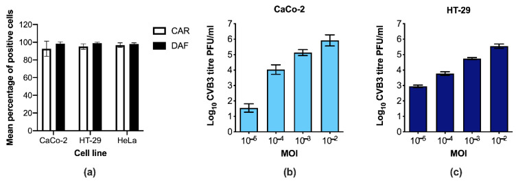
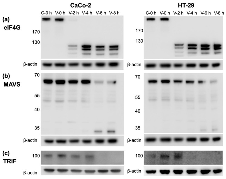
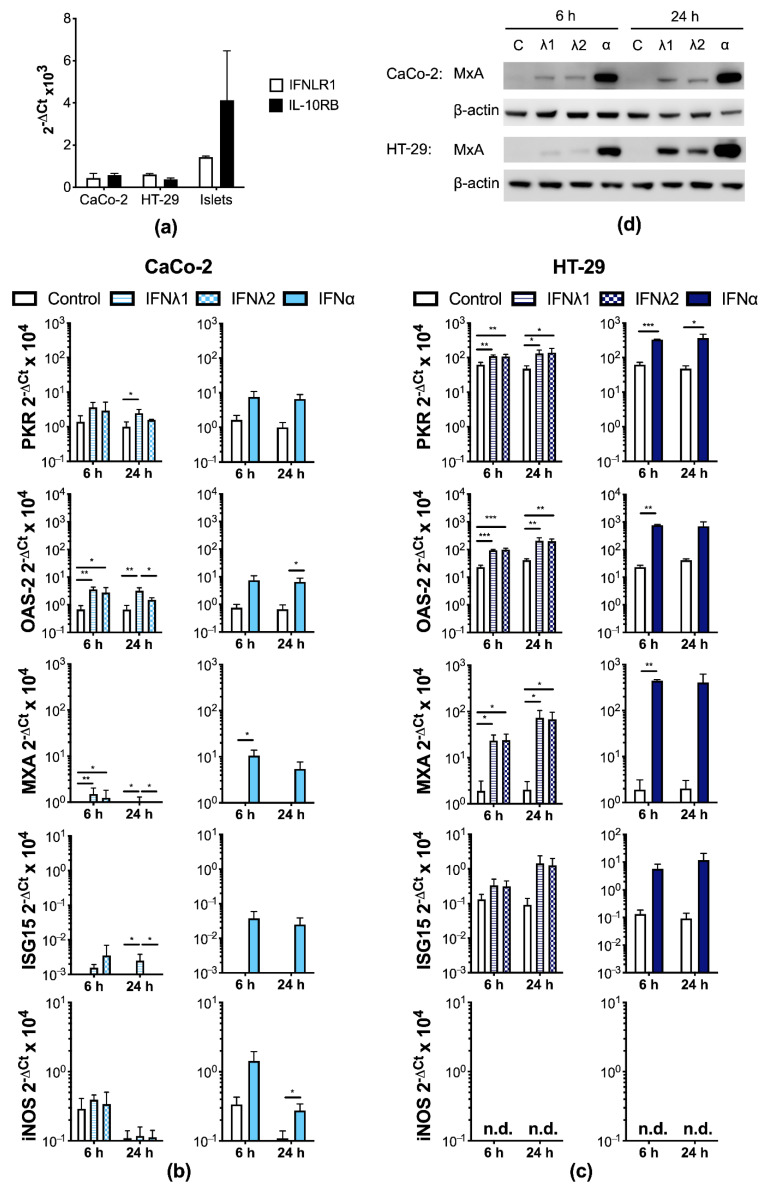
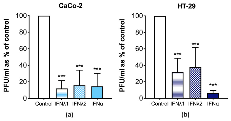
Increasing evidence highlights the importance of the antiviral activities of the type III interferons (IFNλs; IL-28A, IL-28B, IL29, and IFNλ4) in the intestine. However, many viruses have developed strategies to counteract these defense mechanisms by preventing the production of IFNs. Here we use infection models, a clinical virus isolate, and several molecular biology techniques to demonstrate that both type I and III IFNs induce an antiviral state and attenuate Coxsackievirus group B (CVB) replication in human intestinal epithelial cells (IECs). While treatment of IECs with a viral mimic (poly (I:C)) induced a robust expression of both type I and III IFNs, no such up-regulation was observed after CVB infection. The blunted IFN response was paralleled by a reduction in the abundance of proteins involved in the induction of interferon gene transcription, including TIR-domain-containing adapter-inducing interferon-β (TRIF), mitochondrial antiviral-signaling protein (MAVS), and the global protein translation initiator eukaryotic translation initiation factor 4G (eIF4G). Taken together, this study highlights a potent anti-Coxsackieviral effect of both type I and III IFNs in cells located at the primary site of infection. Furthermore, we show for the first time that the production of type I and III IFNs in IECs is blocked by CVBs. These findings suggest that CVBs evade the host immune response in order to successfully infect the intestine.

摘要

越来越多的证据凸显了III型干扰素(IFNλs;IL-28A、IL-28B、IL29和IFNλ4)在肠道中的抗病毒活性的重要性。然而,许多病毒已发展出通过阻止干扰素产生来对抗这些防御机制的策略。在此,我们使用感染模型、一种临床病毒分离株以及多种分子生物学技术来证明I型和III型干扰素均可诱导抗病毒状态并减弱柯萨奇病毒B组(CVB)在人肠道上皮细胞(IECs)中的复制。虽然用病毒模拟物(聚肌苷酸-聚胞苷酸(poly (I:C)))处理IECs可诱导I型和III型干扰素的强烈表达,但CVB感染后未观察到这种上调。干扰素反应减弱的同时,参与干扰素基因转录诱导的蛋白质丰度降低,包括含TIR结构域的衔接蛋白诱导干扰素-β(TRIF)、线粒体抗病毒信号蛋白(MAVS)以及全局蛋白质翻译起始因子真核翻译起始因子4G(eIF4G)。综上所述,本研究凸显了I型和III型干扰素在位于感染原发部位的细胞中具有强大的抗柯萨奇病毒作用。此外,我们首次表明CVB可阻断IECs中I型和III型干扰素的产生。这些发现表明CVB为成功感染肠道而逃避宿主免疫反应。

https://cdn.ncbi.nlm.nih.gov/pmc/blobs/6863/7824802/13d7de529f44/microorganisms-09-00105-g005.jpg
https://cdn.ncbi.nlm.nih.gov/pmc/blobs/6863/7824802/a29029c95ef5/microorganisms-09-00105-g001.jpg
https://cdn.ncbi.nlm.nih.gov/pmc/blobs/6863/7824802/c6bed6a2c46d/microorganisms-09-00105-g002.jpg
https://cdn.ncbi.nlm.nih.gov/pmc/blobs/6863/7824802/52b1ec9b3e88/microorganisms-09-00105-g003.jpg
https://cdn.ncbi.nlm.nih.gov/pmc/blobs/6863/7824802/12c11dd2de11/microorganisms-09-00105-g004a.jpg
https://cdn.ncbi.nlm.nih.gov/pmc/blobs/6863/7824802/13d7de529f44/microorganisms-09-00105-g005.jpg

相似文献

1
Inhibition of Type III Interferon Expression in Intestinal Epithelial Cells-A Strategy Used by Coxsackie B Virus to Evade the Host's Innate Immune Response at the Primary Site of Infection?抑制肠道上皮细胞中III型干扰素的表达——柯萨奇B病毒在感染原发部位逃避宿主固有免疫反应所采用的一种策略?
Microorganisms. 2021 Jan 5;9(1):105. doi: 10.3390/microorganisms9010105.
2
Coxsackievirus counters the host innate immune response by blocking type III interferon expression.柯萨奇病毒通过阻断III型干扰素的表达来对抗宿主的固有免疫反应。
J Gen Virol. 2016 Jun;97(6):1368-1380. doi: 10.1099/jgv.0.000443. Epub 2016 Mar 2.
3
Type III Interferon Restriction by Porcine Epidemic Diarrhea Virus and the Role of Viral Protein nsp1 in IRF1 Signaling.猪流行性腹泻病毒对III型干扰素的限制作用以及病毒蛋白nsp1在IRF1信号传导中的作用
J Virol. 2018 Jan 30;92(4). doi: 10.1128/JVI.01677-17. Print 2018 Feb 15.
4
Type I and Type III Interferons Display Different Dependency on Mitogen-Activated Protein Kinases to Mount an Antiviral State in the Human Gut.I型和III型干扰素在人类肠道中建立抗病毒状态时对丝裂原活化蛋白激酶表现出不同的依赖性。
Front Immunol. 2017 Apr 21;8:459. doi: 10.3389/fimmu.2017.00459. eCollection 2017.
5
Parainfluenza Virus 3 Blocks Antiviral Mediators Downstream of the Interferon Lambda Receptor by Modulating Stat1 Phosphorylation.副流感病毒3型通过调节Stat1磷酸化来阻断干扰素λ受体下游的抗病毒介质。
J Virol. 2015 Dec 30;90(6):2948-58. doi: 10.1128/JVI.02502-15.
6
Type III interferons are expressed by Coxsackievirus-infected human primary hepatocytes and regulate hepatocyte permissiveness to infection.III 型干扰素由感染柯萨奇病毒的人原代肝细胞表达,并调节肝细胞对感染的易感性。
Clin Exp Immunol. 2014 Sep;177(3):687-95. doi: 10.1111/cei.12368.
7
Replication of Human Norovirus RNA in Mammalian Cells Reveals Lack of Interferon Response.人类诺如病毒RNA在哺乳动物细胞中的复制揭示了其缺乏干扰素反应。
J Virol. 2016 Sep 12;90(19):8906-23. doi: 10.1128/JVI.01425-16. Print 2016 Oct 1.
8
Enterovirus 71 Antagonizes Antiviral Effects of Type III Interferon and Evades the Clearance of Intestinal Intraepithelial Lymphocytes.肠道病毒71型拮抗III型干扰素的抗病毒作用并逃避肠道上皮内淋巴细胞的清除。
Front Microbiol. 2022 Feb 1;12:806084. doi: 10.3389/fmicb.2021.806084. eCollection 2021.
9
Susceptibility of primary chicken intestinal epithelial cells for low pathogenic avian influenza virus and velogenic viscerotropic Newcastle disease virus.雏鸡肠道上皮细胞对低致病性禽流感病毒和速发型嗜内脏型新城疫病毒的易感性
Virus Res. 2016 Oct 2;225:50-63. doi: 10.1016/j.virusres.2016.09.001. Epub 2016 Sep 3.
10
Seneca Valley Virus Suppresses Host Type I Interferon Production by Targeting Adaptor Proteins MAVS, TRIF, and TANK for Cleavage.塞内卡谷病毒通过靶向衔接蛋白MAVS、TRIF和TANK进行切割来抑制宿主I型干扰素的产生。
J Virol. 2017 Jul 27;91(16). doi: 10.1128/JVI.00823-17. Print 2017 Aug 15.

引用本文的文献

1
Immunohistochemical Profiling of Interferon-Stimulated Genes in Histopathological Specimens: A Potential Screening Approach for Viral Infections.组织病理学标本中干扰素刺激基因的免疫组织化学分析:一种潜在的病毒感染筛查方法。
APMIS. 2025 Jun;133(6):e70034. doi: 10.1111/apm.70034.
2
Baicalein suppresses Coxsackievirus B3 replication by inhibiting caspase-1 and viral protease 2A.黄芩素通过抑制半胱天冬酶-1 和病毒蛋白酶 2A 来抑制柯萨奇病毒 B3 的复制。
Virol Sin. 2024 Aug;39(4):685-693. doi: 10.1016/j.virs.2024.07.003. Epub 2024 Jul 16.
3
Role of type-I and type-III interferons in gastrointestinal homeostasis and pathogenesis.

本文引用的文献

1
The TLR3/IRF1/Type III IFN Axis Facilitates Antiviral Responses against Enterovirus Infections in the Intestine.TLR3/IRF1/III 型 IFN 轴促进肠道对抗肠道病毒感染的抗病毒反应。
mBio. 2020 Nov 17;11(6):e02540-20. doi: 10.1128/mBio.02540-20.
2
Defining the proteolytic landscape during enterovirus infection.定义肠道病毒感染期间的蛋白水解全景。
PLoS Pathog. 2020 Sep 30;16(9):e1008927. doi: 10.1371/journal.ppat.1008927. eCollection 2020 Sep.
3
Critical Role of Type III Interferon in Controlling SARS-CoV-2 Infection in Human Intestinal Epithelial Cells.
I 型和 III 型干扰素在胃肠道稳态和发病机制中的作用。
Curr Opin Immunol. 2024 Feb;86:102412. doi: 10.1016/j.coi.2024.102412. Epub 2024 Mar 21.
4
Current Landscape of IFN-λ: Induction, Inhibition, and Potential Clinical Applications to Treat Respiratory Viral Infections.IFN-λ 的现状:诱导、抑制及其治疗呼吸道病毒感染的潜在临床应用。
Immunohorizons. 2023 Apr 1;7(4):265-272. doi: 10.4049/immunohorizons.2200010.
5
Coxsackievirus Infection and Associated Diseases.柯萨奇病毒感染及相关疾病
Microorganisms. 2022 Aug 4;10(8):1566. doi: 10.3390/microorganisms10081566.
6
Effect of cannabidiol on apoptosis and cellular interferon and interferon-stimulated gene responses to the SARS-CoV-2 genes ORF8, ORF10 and M protein.大麻二酚对 SARS-CoV-2 基因 ORF8、ORF10 和 M 蛋白的细胞凋亡和细胞干扰素及干扰素刺激基因反应的影响。
Life Sci. 2022 Jul 15;301:120624. doi: 10.1016/j.lfs.2022.120624. Epub 2022 May 11.
7
Virus Infection Is an Instigator of Intestinal Dysbiosis Leading to Type 1 Diabetes.病毒感染是导致 1 型糖尿病的肠道菌群失调的诱因。
Front Immunol. 2021 Oct 15;12:751337. doi: 10.3389/fimmu.2021.751337. eCollection 2021.
8
Viruses and Type 1 Diabetes: From Enteroviruses to the Virome.病毒与1型糖尿病:从肠道病毒到病毒组
Microorganisms. 2021 Jul 16;9(7):1519. doi: 10.3390/microorganisms9071519.
9
Immune Transcriptome of Cells Infected with Enterovirus Strains Obtained from Cases of Autoimmune Thyroid Disease.从自身免疫性甲状腺疾病病例中获得的肠道病毒株感染细胞的免疫转录组
Microorganisms. 2021 Apr 19;9(4):876. doi: 10.3390/microorganisms9040876.
III 型干扰素在控制 SARS-CoV-2 感染人肠上皮细胞中的关键作用。
Cell Rep. 2020 Jul 7;32(1):107863. doi: 10.1016/j.celrep.2020.107863. Epub 2020 Jun 19.
4
Type III interferons: Balancing tissue tolerance and resistance to pathogen invasion.III 型干扰素:平衡组织耐受和抵御病原体入侵。
J Exp Med. 2020 Jan 6;217(1). doi: 10.1084/jem.20190295.
5
Prospective virome analyses in young children at increased genetic risk for type 1 diabetes.对 1 型糖尿病遗传风险增加的幼儿进行前瞻性病毒组分析。
Nat Med. 2019 Dec;25(12):1865-1872. doi: 10.1038/s41591-019-0667-0. Epub 2019 Dec 2.
6
Genetic and Environmental Interaction in Type 1 Diabetes: a Relationship Between Genetic Risk Alleles and Molecular Traits of Enterovirus Infection?1 型糖尿病的遗传与环境交互作用:遗传风险等位基因与肠道病毒感染的分子特征之间的关系?
Curr Diab Rep. 2019 Aug 10;19(9):82. doi: 10.1007/s11892-019-1192-8.
7
Interferon-λ orchestrates innate and adaptive mucosal immune responses.干扰素-λ 调控先天和适应性黏膜免疫应答。
Nat Rev Immunol. 2019 Oct;19(10):614-625. doi: 10.1038/s41577-019-0182-z. Epub 2019 Jun 14.
8
Enteroviruses: A Gut-Wrenching Game of Entry, Detection, and Evasion.肠道病毒:进入、检测和逃避的惊心动魄的游戏。
Viruses. 2019 May 21;11(5):460. doi: 10.3390/v11050460.
9
Shared and Distinct Functions of Type I and Type III Interferons.I 型和 III 型干扰素的共有和独特功能。
Immunity. 2019 Apr 16;50(4):907-923. doi: 10.1016/j.immuni.2019.03.025.
10
Essential Role of Enterovirus 2A Protease in Counteracting Stress Granule Formation and the Induction of Type I Interferon.肠病毒 2A 蛋白酶在对抗应激颗粒形成和诱导 I 型干扰素中的重要作用。
J Virol. 2019 May 1;93(10). doi: 10.1128/JVI.00222-19. Print 2019 May 15.