Department of Gastroenterology, St. Luke's International Hospital, 9-1 Akashicho, Chuo-ku, Tokyo, 104-8560, Japan.
BMC Gastroenterol. 2021 Jan 19;21(1):29. doi: 10.1186/s12876-021-01607-w.
Mucosal Schwann cell hamartomas are rare neurogenic tumors which present most commonly in the distal colon. They are usually discovered as small, single polyps in asymptomatic patients.
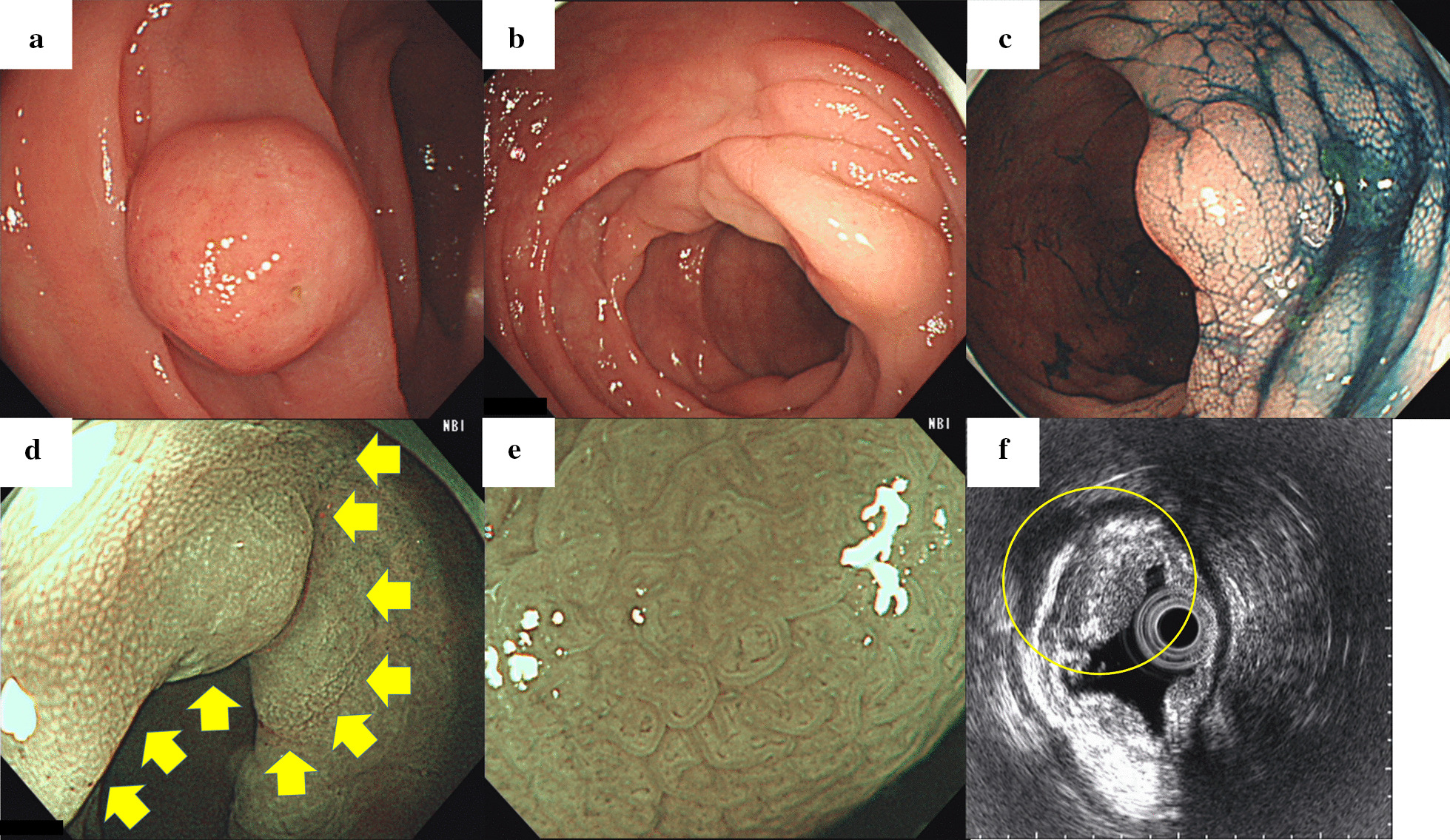
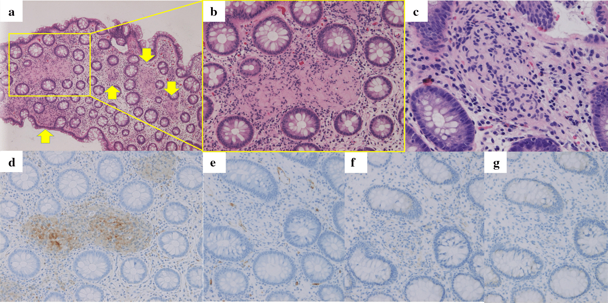
An asymptomatic 64-year-old man was referred to us after a 12 mm subepithelial lesion was discovered incidentally on screening colonoscopy. Follow-up colonoscopy conducted 2 months later revealed that the tumor had disappeared, leaving multiple edematous, submucosal tumor-like elevations presenting as skip lesions throughout the sigmoid colon. Lesions had elongated pits on magnifying endoscopy and were limited to the first layer on endoscopic ultrasound. Biopsies revealed unclearly delineated foci of spindle-shaped cells limited to the lamina propria. On immunohistochemistry, all lesions were positive for S-100 protein and negative for CD34, CD56, and neurofilament protein. The patient was diagnosed with multiple mucosal Schwann cell hamartomas of the sigmoid colon. He remains asymptomatic after 18 months of follow-up, but the lesions have also remained unchanged.
We report a case of multiple non-polypoid mucosal Schwann cell hamartomas. Endoscopic findings may assist in the differential diagnosis, particularly when presenting as non-polypoid, submucosal tumor-like lesions.
黏膜 Schwann 细胞错构瘤是一种罕见的神经源性肿瘤,最常发生于远端结肠。它们通常在无症状患者中作为单个小息肉被发现。
一位 64 岁无症状男性因筛查性结肠镜检查偶然发现 12mm 黏膜下病变而被转介至我们处。2 个月后进行的随访结肠镜检查显示,肿瘤已消失,而遗留多发黏膜下、类似肿瘤样隆起的水肿性病变,呈跳跃性分布于乙状结肠。放大内镜下病变可见长形凹陷,且仅在超声内镜的第一层发现病变。活检显示固有层内界限不清的梭形细胞灶。免疫组化显示,所有病变均 S-100 蛋白阳性,CD34、CD56 和神经丝蛋白阴性。患者被诊断为乙状结肠多发黏膜 Schwann 细胞错构瘤。随访 18 个月后,患者仍无症状,但病变也未改变。
我们报告了一例多发非息肉样黏膜 Schwann 细胞错构瘤。内镜表现有助于鉴别诊断,尤其是当表现为非息肉样、黏膜下类似肿瘤样病变时。