Lu Yunping, Li Lingyu, Chen Hui, Jing Xinying, Wang Min, Ge Lihua, Yang Jing, Zhang Min, Tang Xiaofei
Beijing Institute of Dental Research, Beijing Key Laboratory, Beijing Stomatological Hospital & School of Stomatology, Capital Medical University, Beijing 100050, People's Republic of China.
Onco Targets Ther. 2021 Jan 12;14:239-251. doi: 10.2147/OTT.S284182. eCollection 2021.
Cellular senescence is a physiological phenomenon by which cells irreversibly lose their proliferative potential. It is not clear whether senescent cells are related to malignant transformation in oral precancerous lesions. The role of peroxiredoxin1 (Prx1)-induced cell senescence in OLK malignant transformation has not been reported. The aim of this study is to investigate the role and mechanism of cell senescence in oral carcinogenesis.
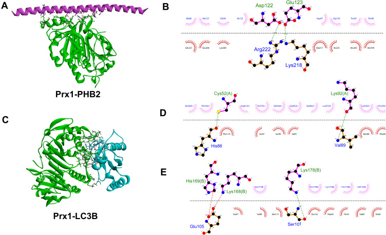
In this study, 4-nitro-quinoline-1-oxide (4NQO) induced tongue carcinogenesis model in Prx1 and Prx1 mice and dysplastic oral keratinocyte (DOK) were used. Prx1 knockdown DOK cells were harvested with shRNA injection, and cell senescence was detected via the senescence-associated β-galactosidase (SA β-gal) assay. The senescence and mitophagy-related proteins were observed by immunohistochemistry (IHC), Western blot and qRT-PCR. The binding of Prx1 with prohibitin 2 (PHB2) and light chain 3 (LC3) was predicted via ZDOCK and measured in mice by Duolink analysis.
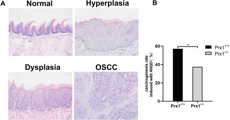
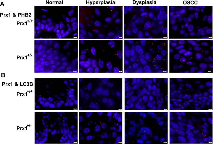
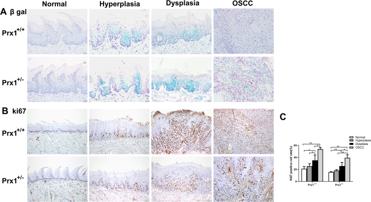
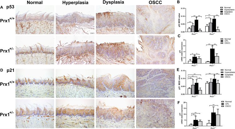
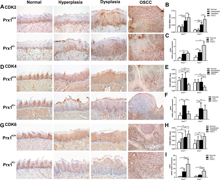
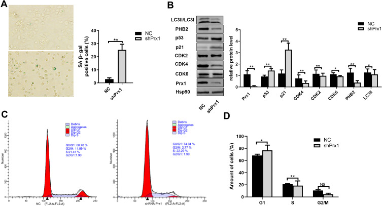
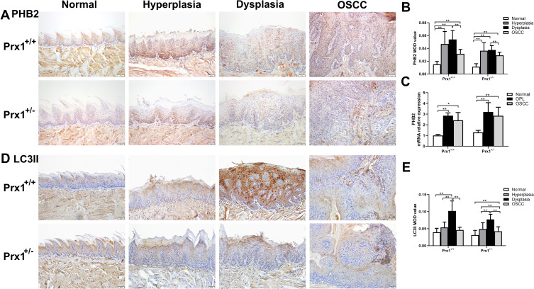
Histologically, 4NQO treatment induced epithelial hyperplasia, dysplasia (mild, moderate and severe), carcinomas in situ and oral squamous cell carcinoma (OSCC) in mouse tongue mucosa. The malignant transformation rate in Prx1 mice (37.5%) was significantly lower compared with Prx1 mice (57.1%). In Prx1 mice, a higher number of senescent cells and greater expression of p53 and p21 were observed in hyperplastic and dysplastic tongue tissues when compared with those in OSCC tissues. Prx1 knockdown induced a greater number of senescent cells in hyperplastic tissues, and DOK cells accompanied cell cycle arrest at the G1 phase and PHB2/LC3II downregulation. Prx1 was predicted to dock with PHB2 and LC3 via ZDOCK, and the interactions were confirmed by in situ Duolink analysis.
Prx1 silencing inhibits the oral carcinogenesis by inducing cell senescence dependent on mitophagy.
细胞衰老一种细胞不可逆地丧失增殖潜能的生理现象。目前尚不清楚衰老细胞是否与口腔癌前病变的恶性转化有关。过氧化物酶1(Prx1)诱导的细胞衰老在口腔黏膜下纤维化(OLK)恶性转化中的作用尚未见报道。本研究旨在探讨细胞衰老在口腔癌发生中的作用及机制。
在本研究中,使用4-硝基喹啉-1-氧化物(4NQO)诱导Prx1和Prx1小鼠的舌癌发生模型以及发育异常的口腔角质形成细胞(DOK)。通过注射shRNA收获Prx1敲低的DOK细胞,并通过衰老相关β-半乳糖苷酶(SA β-gal)检测法检测细胞衰老。通过免疫组织化学(IHC)、蛋白质免疫印迹法和qRT-PCR观察衰老和线粒体自噬相关蛋白。通过ZDOCK预测Prx1与抗增殖蛋白2(PHB2)和微管相关蛋白1轻链3(LC3)的结合,并通过Duolink分析在小鼠中进行测量。
组织学上,4NQO处理诱导小鼠舌黏膜上皮增生、发育异常(轻度、中度和重度)、原位癌和口腔鳞状细胞癌(OSCC)。与Prx1小鼠(57.1%)相比,Prx1小鼠的恶性转化率(37.5%)显著降低。在Prx1小鼠中,与OSCC组织相比,在增生性和发育异常的舌组织中观察到更多的衰老细胞以及更高的p53和p21表达。Prx1敲低在增生组织中诱导更多的衰老细胞,并且DOK细胞伴随细胞周期停滞在G1期以及PHB2/LC3II下调。通过ZDOCK预测Prx1与PHB2和LC3对接,并且通过原位Duolink分析证实了这种相互作用。
Prx1沉默通过诱导依赖线粒体自噬的细胞衰老来抑制口腔癌发生。