Department of Neurology, Medical College of Wisconsin, Milwaukee, Wisconsin, USA.
Department of Dermatology, Medical College of Wisconsin, Milwaukee, Wisconsin, USA.
Photobiomodul Photomed Laser Surg. 2021 Jun;39(6):411-417. doi: 10.1089/photob.2020.4957. Epub 2021 Jan 20.
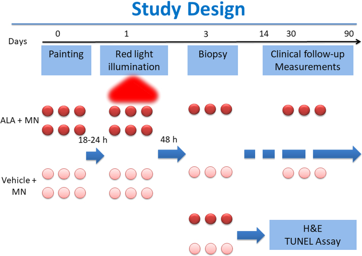
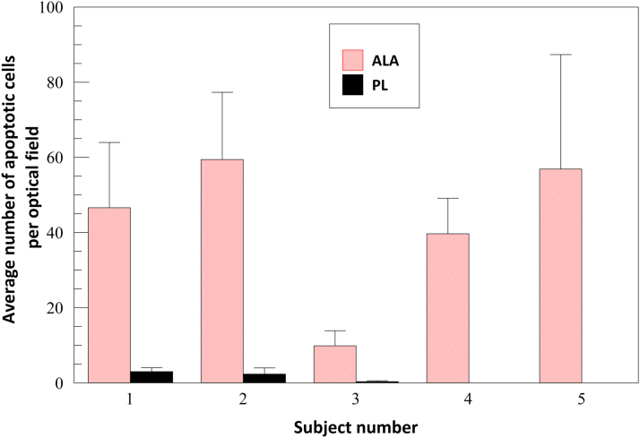
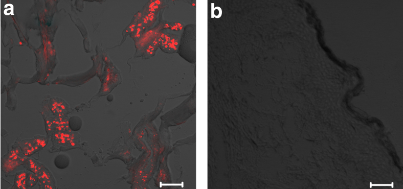
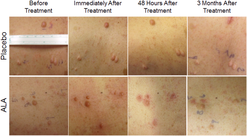
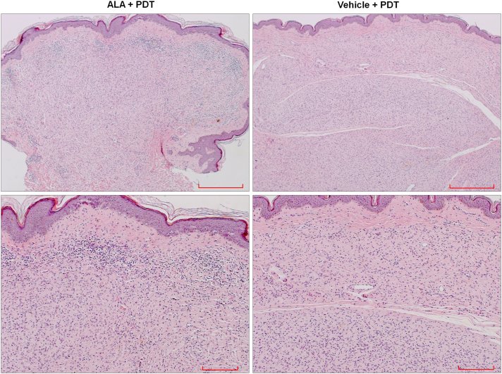
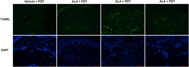
Neurofibromatosis type 1 (NF1) has no current effective treatments beyond surgery. Topical photodynamic therapy (PDT) has the potential to provide a less invasive treatment modality. Based on murine data, we hypothesized PDT could be used for the treatment of cutaneous neurofibromas (cNF). We conducted a phase I trial to examine absorption and conversion of topical aminolevulinic acid (ALA) in cNF and determine safety in a dose escalation study. ALA or control vehicle was applied to neurofibromas through microneedle-assisted delivery ( = 4) and excised specimens were examined 24 h later for protoporphyrin IX fluorescence. Fluorescence was detected in the tumors at 304 ± 94 U/μm, while adjacent paralesional normal skin and vehicle-treated tumors showed no fluorescence ( < 0.0001). Subsequently, neurofibromas ( = 27) were treated with ALA and irradiated with 633 nm red light 18 h later, at escalating dosages of 50 and 100 mJ/cm. Maximum tolerable dose was established at 100 mJ/cm. Light microscopy study of tumors biopsied 48 h after PDT (ALA = 14 and vehicle = 4) showed mixed inflammatory infiltrate in the ALA, but not in the vehicle-treated tumors or perilesional normal skin. TUNEL evaluation showed 42.5 ± 19.9 apoptotic cells per visual field for ALA-treated and 1.1 ± 1.4 for vehicle-treated tumors ( = 0.002). In the first reported clinical trial of PDT for NF1, PDT targeted neurofibromas specifically, and may offer a normal tissue-sparing treatment modality in the future. This study is registered at Clintrials.gov (NCT01682811).
神经纤维瘤病 1 型(NF1)除手术外尚无有效的治疗方法。局部光动力疗法(PDT)有可能提供一种微创的治疗方式。基于鼠类数据,我们假设 PDT 可用于治疗皮肤神经纤维瘤(cNF)。我们进行了一项 I 期临床试验,以检查 cNF 中局部氨茴酸(ALA)的吸收和转化,并在剂量递增研究中评估安全性。ALA 或对照载体通过微针辅助输送应用于神经纤维瘤( = 4),并在 24 小时后切除标本检查原卟啉 IX 荧光。肿瘤中检测到 304 ± 94 U/μm 的荧光,而相邻旁皮损正常皮肤和对照载体处理的肿瘤无荧光( < 0.0001)。随后,18 小时后,用 633nm 红光照射神经纤维瘤( = 27),ALA 剂量递增至 50 和 100 mJ/cm。最大耐受剂量确定为 100 mJ/cm。PDT 后 48 小时活检肿瘤的光镜研究(ALA = 14,载体 = 4)显示,ALA 中的混合炎症浸润,而在载体处理的肿瘤或旁皮损正常皮肤中则没有。TUNEL 评估显示,ALA 治疗组每个视野有 42.5 ± 19.9 个凋亡细胞,而载体治疗组有 1.1 ± 1.4 个凋亡细胞( = 0.002)。在 NF1 的 PDT 首次临床报告中,PDT 特异性靶向神经纤维瘤,将来可能提供一种对正常组织无损伤的治疗方式。这项研究在 Clintrials.gov 注册(NCT01682811)。