Department of Diagnostic and Interventional Radiology, School of Medicine & Klinikum rechts der Isar, Technical University of Munich, Ismaningerstr. 22, 81675, Munich, Germany.
Philips CT Clinical Science, Hamburg, Germany.
Eur Radiol. 2021 Aug;31(8):6193-6199. doi: 10.1007/s00330-020-07677-w. Epub 2021 Jan 20.
Determination of coronary artery calcium scoring (CACS) in non-contrast computed tomography (CT) images has been shown to be an important prognostic factor in coronary artery disease (CAD). The objective of this study was to evaluate the accuracy of CACS from virtual non-contrast (VNC) imaging generated from spectral data in comparison to standard (true) non-contrast (TNC) imaging in a representative patient cohort with clinically approved software.
One hundred three patients referred to coronary CTA with suspicion of CAD were investigated on a dual-layer spectral detector CT (SDCT) scanner. CACS was calculated from both TNC and VNC images by software certified for medical use. Patients with a CACS of 0 were excluded from analysis.
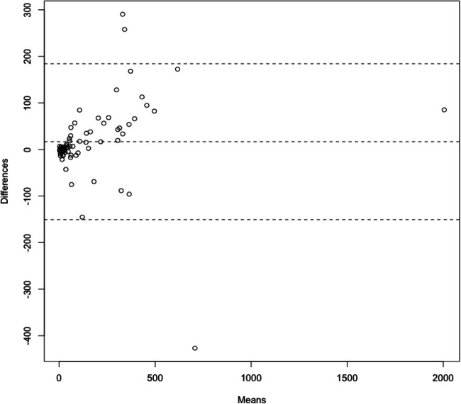
The mean age of the study population was 61 ± 11 years with 48 male patients (67%). Inter-quartile range of clinical CACS was 22-282. Correlation of measured CACS from true- and VNC images was high (0.95); p < 0.001. The slope was 3.83, indicating an underestimation of VNC CACS compared to TNC CACS by that factor. Visual analysis of the Bland-Altman plot of CACS showed good accordance with both methods after correction of VNC CACS by the abovementioned factor.
In clinical diagnostics of CAD, the determination of CACS is feasible using VNC images generated from spectral data obtained on a dual-layer spectral detector CT. When multiplied by a correction factor, results were in good agreement with the standard technique. This could enable radiation dose reductions by obviating the need for native scans typically used for CACS.
• Calcium scoring is feasible from contrast-enhanced CT images using a dual-layer spectral detector CT scanner. • When multiplied by a correction factor, calcium scoring from virtual non-contrast images shows good agreement with the standard technique. • Omitting native scans for calcium scoring could enable radiation dose reduction.
在非对比计算机断层扫描(CT)图像中进行冠状动脉钙评分(CACS)已被证明是冠状动脉疾病(CAD)的一个重要预后因素。本研究的目的是评估在具有临床批准软件的代表性患者队列中,从光谱数据生成的虚拟非对比(VNC)成像与标准(真实)非对比(TNC)成像相比,CACS 的准确性。
对怀疑患有 CAD 的 103 名患者进行双层光谱探测器 CT(SDCT)扫描仪冠状动脉 CTA 检查。使用经医疗认证的软件从 TNC 和 VNC 图像计算 CACS。将 CACS 为 0 的患者排除在分析之外。
研究人群的平均年龄为 61±11 岁,其中 48 名男性患者(67%)。临床 CACS 的四分位间距为 22-282。真实和 VNC 图像测量的 CACS 之间存在高度相关性(0.95);p<0.001。斜率为 3.83,表明与 TNC CACS 相比,VNC CACS 低估了该因素的 3.83 倍。经上述因素校正后,Bland-Altman 图的 CACS 视觉分析表明,两种方法的一致性良好。
在 CAD 的临床诊断中,使用双层光谱探测器 CT 获得的光谱数据生成的 VNC 图像可实现 CACS 的测定。当乘以校正因子时,结果与标准技术非常吻合。这可以通过省去通常用于 CACS 的原始扫描来减少辐射剂量。
使用双层光谱探测器 CT 扫描仪可以从增强 CT 图像中进行钙评分。
当乘以校正因子时,虚拟非对比图像的钙评分与标准技术具有良好的一致性。
省略钙评分的原始扫描可以减少辐射剂量。