Al-Hindi Mariam, Al-Fotawi Randa, Al-Tamimi Abdulaziz, Khalil Osama, Al-Osaimi Naif, Al-Ghamdi Khalid, Heji Khloud
Oral and Maxillofacial Surgery Dept, Dental Collage, King Saud University, Saudi Arabia.
Oral and Maxillofacial Surgery Dept, Dental Collage, King Saud University, Saudi Arabia.
Int J Surg Case Rep. 2021 Feb;79:255-262. doi: 10.1016/j.ijscr.2021.01.040. Epub 2021 Jan 15.
Hypothyroidism reduces the recruitment, maturation, and activity of bone cells, decreasing bone resorption and formation. Several investigations have reported that T4 replacement therapy is associated with a significant decrease in bone mineral density in various skeletal parts, while others have failed to corroborate these results. The present study describes both a literature review and our own experience with dental implants in patients with hypothyroidism undergoing T4 replacement therapy.
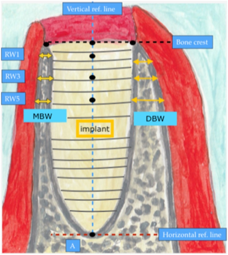
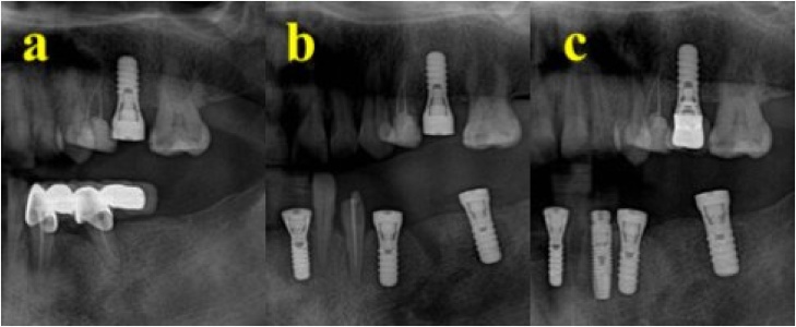
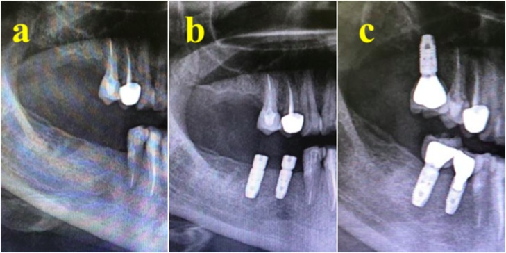
The study included two parts: a literature review and case series. The literature review included 12 articles documenting the success rate of osseointegrated dental implants in patients with hypothyroidism. The clinical cases were chosen from King Saud University Dental College, Riyadh. The patients' identity was only available to the main researcher. The inclusion criteria for the clinical cases were: T4-treated hypothyroidism, age 20-60 years, and use of dental implants with a follow-up period of 6-12 months after loading. The exclusion criteria were: any other medical condition alongside hypothyroidism, syndromic hypothyroidism, pregnancy, current smoking, bruxism, and hypothyroidism caused by surgical excision combined with radiotherapy. The following parameters were assessed: insertion torque (IT), crestal bone height (CBH), mesial bone width (MBW), and distal bone width (DBW) at different time points, probing depth, and signs of infection.
Seventeen dental implants placed in patients with T4-treated hypothyroidism showed median IT success (42.4 N⋅cm; range: 35-45 N⋅cm). The median crestal bone loss was measured at 6-12 months after loading was 0.6 mm (range: 0.5-0.7. mm). Conversely, the median bone loss differences in MBW and DBW at 6-12 months after loading were 0.3 mm and 0.2 mm, respectively.
After a 1-year follow-up, patients with T4-treated hypothyroidism who had received dental implants fulfilled the criteria for successful implants.
甲状腺功能减退会降低骨细胞的募集、成熟和活性,减少骨吸收和形成。多项研究报告称,甲状腺素替代疗法与各个骨骼部位骨密度的显著降低有关,而其他研究则未能证实这些结果。本研究既描述了文献综述,也阐述了我们自身在接受甲状腺素替代疗法的甲状腺功能减退患者中进行牙种植的经验。
该研究包括两个部分:文献综述和病例系列。文献综述纳入了12篇记录甲状腺功能减退患者骨结合牙种植成功率的文章。临床病例选自利雅得的沙特国王大学牙科学院。只有主要研究者知道患者的身份。临床病例的纳入标准为:接受甲状腺素治疗的甲状腺功能减退症、年龄20至60岁、使用牙种植体且负重后随访6至12个月。排除标准为:除甲状腺功能减退症外的任何其他疾病、综合征性甲状腺功能减退症、妊娠、当前吸烟、磨牙症以及手术切除联合放疗导致的甲状腺功能减退症。评估了以下参数:不同时间点的植入扭矩(IT)、嵴顶骨高度(CBH)、近中骨宽度(MBW)和远中骨宽度(DBW)、探诊深度以及感染迹象。
为接受甲状腺素治疗的甲状腺功能减退患者植入的17颗牙种植体显示IT成功率中位数为(42.4 N·cm;范围:35至45 N·cm)。负重后6至12个月测量的嵴顶骨丢失中位数为0.6 mm(范围:0.5至0.7 mm)。相反,负重后6至12个月MBW和DBW的骨丢失差异中位数分别为0.3 mm和0.2 mm。
经过1年的随访,接受牙种植的接受甲状腺素治疗的甲状腺功能减退患者符合种植成功的标准。