Pagiola Igor, Amaral Bruno, Saito Celso, Nalli Darcio, Carrete Junior Henrique, Frudit Michel
Department of Interventional Neuroradiology, Universidade Federal de São Paulo, São Paulo SP, Brazil.
Department of Interventional Neuroradiology, Hospital Estadual Central, VitÓria ES, Brazil.
J Cerebrovasc Endovasc Neurosurg. 2021 Mar;23(1):60-63. doi: 10.7461/jcen.2021.E2020.05.001. Epub 2021 Jan 26.
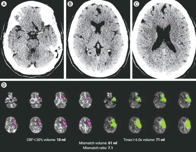
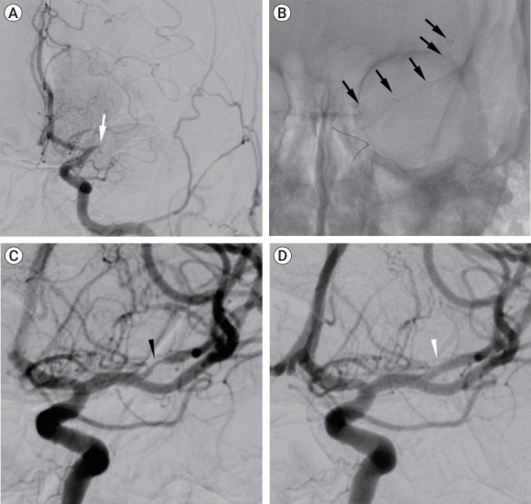
Here we describe a successful mechanical thrombectomy (MT) for acute large vessel occlusion in stroke treatment with one passage (thrombolysis in cerebral infarction, TICI 3). Immediately after the withdrawing of the stent retriever, a narrowing of the middle cerebral artery was diagnosed. The rate of vasospasms during this procedure can be as higher as 41% (range from 6-41%). Here we describe our protocol when a narrowing of the artery is visualized after a stent retriever is withdrawn. A patient presented in our emergency room with National Institute of Health Stroke Scale (NIHSS) of 21, Alberta Stroke Program Early CT Score (ASPECTS) 8, computed tomography angiography revealed occlusion of the M1 segment and MT was indicated. One passage TICI Ⅲ was achieved. After that, the image showed a narrowing of the artery. We present one case of a spasm after stent retriever technique for MT, we injected vasodilator and the artery became normal in a few minutes differentiating between atheromatous stenosis and vasospasm. We present a technical note that can help to make the differentiation of vasospasm or atheromatous disease after MT with the stent retriever technique.
在此,我们描述了一例在卒中治疗中通过一次操作成功进行机械取栓(MT)治疗急性大血管闭塞的病例(脑梗死溶栓,TICI 3级)。在取出支架取栓器后,立即诊断出大脑中动脉狭窄。在此过程中血管痉挛的发生率可能高达41%(范围为6%-41%)。在此,我们描述当取出支架取栓器后发现动脉狭窄时我们的处理方案。一名患者因美国国立卫生研究院卒中量表(NIHSS)评分为21分、阿尔伯塔卒中项目早期CT评分(ASPECTS)为8分而就诊于我们的急诊室,计算机断层血管造影显示M1段闭塞,遂行MT治疗。实现了一次操作达到TICIⅢ级。此后,影像显示动脉狭窄。我们呈现了一例采用支架取栓器技术进行MT后发生痉挛的病例,我们注射了血管扩张剂,动脉在几分钟内恢复正常,从而区分了动脉粥样硬化性狭窄和血管痉挛。我们提供了一份技术说明,有助于在采用支架取栓器技术进行MT后区分血管痉挛或动脉粥样硬化性疾病。