Department of Radiological Sciences, Medical Sciences I, B-140, University of California, Irvine, CA, 92697, USA.
Int J Cardiovasc Imaging. 2021 May;37(5):1767-1779. doi: 10.1007/s10554-020-02130-x. Epub 2021 Jan 27.
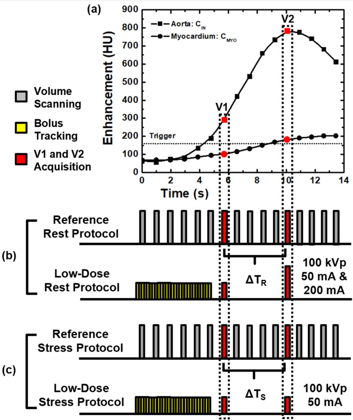
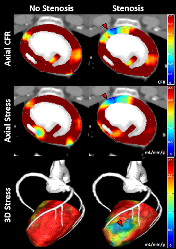
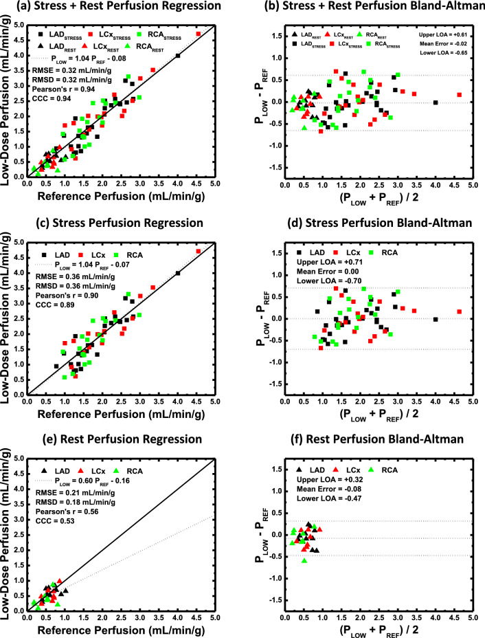
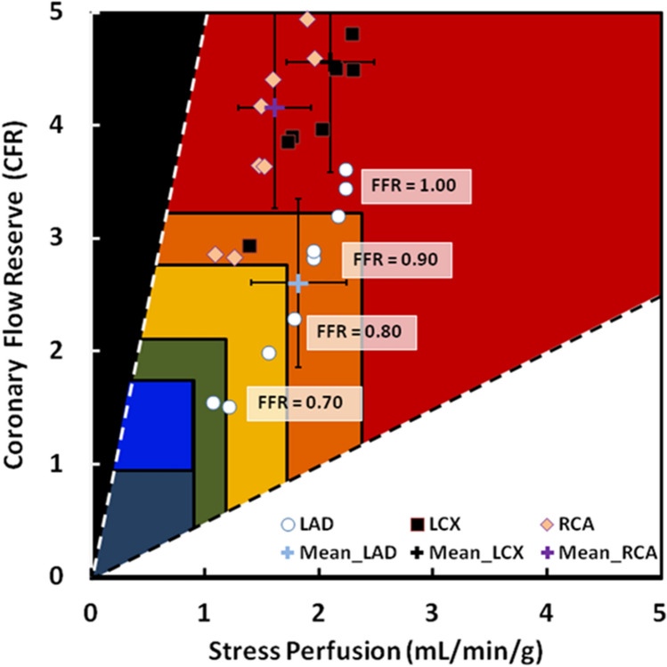
Morphological and physiological assessment of coronary artery disease (CAD) is necessary for proper stratification of CAD risk. The objective was to evaluate a low-dose cardiac CT technique that combines morphological and physiological assessment of CAD. The low-dose technique was evaluated in twelve swine, where three of the twelve had coronary balloon stenosis. The technique consisted of rest perfusion measurement combined with angiography followed by stress perfusion measurement, where the ratio of stress to rest was used to derive coronary flow reserve (CFR). The technique only required two volume scans for perfusion measurement in mL/min/g; hence, four volume scans were acquired in total; two for rest with angiography and two for stress. All rest, stress, and CFR measurements were compared to a previously validated reference technique that employed 20 consecutive volume scans for rest perfusion measurement combined with angiography, and stress perfusion measurement, respectively. The 32 cm diameter volumetric CT dose index ([Formula: see text]) and size-specific dose estimate (SSDE) of the low-dose technique were also recorded. All low-dose perfusion measurements (P) in mL/min/g were related to reference perfusion measurements (P) through regression by P = 1.04 P - 0.08 (r = 0.94, RMSE = 0.32 mL/min/g). The [Formula: see text] and SSDE of the low-dose cardiac CT technique were 8.05 mGy and 12.80 mGy respectively, corresponding to an estimated effective dose and size-specific effective dose of 1.8 and 2.87 mSv, respectively. Combined morphological and physiological assessment of coronary artery disease is feasible using a low-dose cardiac CT technique.
评估冠状动脉疾病(CAD)的形态和生理功能对于适当分层 CAD 风险非常必要。本研究旨在评估一种结合 CAD 形态和生理功能评估的低剂量心脏 CT 技术。该低剂量技术在 12 头猪中进行了评估,其中 12 头中有 3 头存在冠状动脉球囊狭窄。该技术包括静息灌注测量与血管造影相结合,然后进行负荷灌注测量,其中负荷与静息的比值用于得出冠状动脉血流储备(CFR)。该技术仅需两次容积扫描即可获得灌注测量值(mL/min/g);因此,总共采集了四次容积扫描;两次用于静息和血管造影,两次用于负荷。所有静息、负荷和 CFR 测量值均与先前验证的参考技术进行比较,该技术使用 20 次连续容积扫描分别用于静息灌注测量与血管造影,以及负荷灌注测量。还记录了低剂量技术的 32cm 直径容积 CT 剂量指数([Formula: see text])和体积剂量长度乘积特异性剂量估计值(SSDE)。所有低剂量灌注测量值(P)均以 mL/min/g 为单位,通过回归与参考灌注测量值(P)相关,P=1.04 P-0.08(r=0.94,RMSE=0.32 mL/min/g)。低剂量心脏 CT 技术的[Formula: see text]和 SSDE 分别为 8.05 mGy 和 12.80 mGy,分别相当于估计的有效剂量和体积剂量长度乘积特异性有效剂量 1.8 和 2.87 mSv。使用低剂量心脏 CT 技术可以对冠状动脉疾病进行形态和生理联合评估。