Department of Ophthalmology & Visual Sciences, Medical College of Wisconsin, Milwaukee, WI, USA.
Department of Cell Biology, Neurobiology and Anatomy, Medical College of Wisconsin, Milwaukee, WI, USA.
Transl Vis Sci Technol. 2021 Jan 7;10(1):11. doi: 10.1167/tvst.10.1.11. eCollection 2021 Jan.
To determine whether artifacts in optical coherence tomography (OCT) images are associated with the success or failure of adaptive optics scanning light ophthalmoscopy (AOSLO) imaging in subjects with achromatopsia (ACHM).
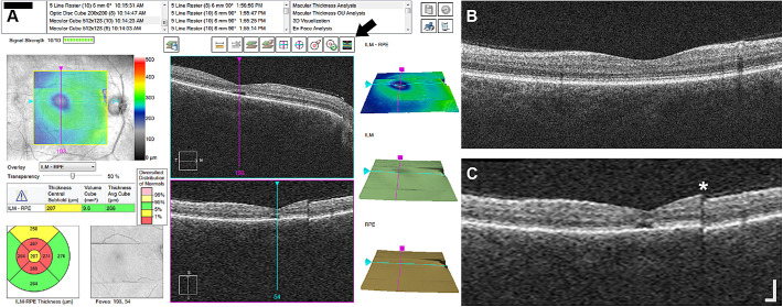
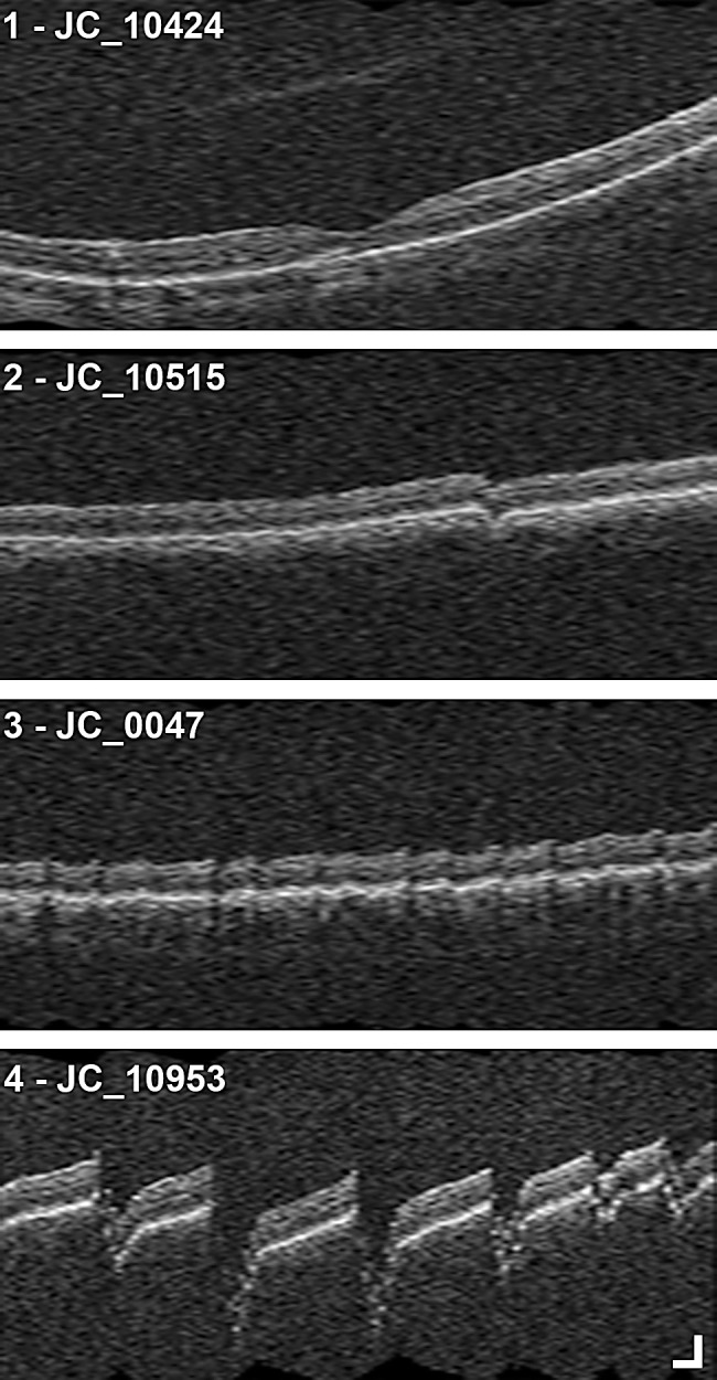
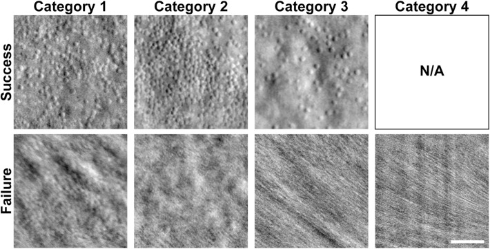
Previously acquired OCT and non-confocal, split-detector AOSLO images from one eye of 66 subjects with genetically confirmed achromatopsia (15 and 51 ) were reviewed along with best-corrected visual acuity (BCVA) and axial length. OCT artifacts in interpolated vertical volumes from CIRRUS macular cubes were divided into four categories: (1) none or minimal, (2) clear and low frequency, (3) low amplitude and high frequency, and (4) high amplitude and high frequency. Each vertical volume was assessed once by two observers. AOSLO success was defined as sufficient image quality in split-detector images at the fovea to assess cone quantity.
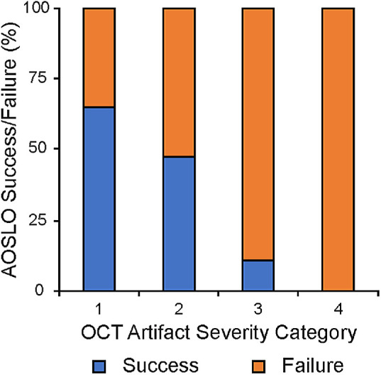
There was excellent agreement between the two observers for assessing OCT artifact severity category (weighted kappa = 0.88). Overall, AOSLO success was 47%. For subjects with OCT artifact severity category 1, AOSLO success was 65%; for category 2, 47%; for category 3, 11%; and for category 4, 0%. There was a significant association between OCT artifact severity category and AOSLO success ( = 0.0002). Neither BCVA nor axial length was associated with AOSLO success ( = 0.07 and = 0.75, respectively).
Artifacts in OCT volumes are associated with AOSLO success in ACHM. Subjects with less severe OCT artifacts are more likely to be good candidates for AOSLO imaging, whereas AOSLO was successful in only 7% of subjects with category 3 or 4 OCT artifacts. These results may be useful in guiding patient selection for AOSLO imaging.
Using OCT to prescreen patients could be a valuable tool for clinical trials that utilize AOSLO to reduce costs and decrease patient testing burden.
确定光学相干断层扫描(OCT)图像中的伪影是否与患有色盲(ACHM)的受试者自适应光学扫描检眼镜(AOSLO)成像的成功或失败相关。
回顾了 66 名经基因证实的色盲(ACHM)患者一只眼的先前获得的 OCT 和非共焦、分束探测器 AOSLO 图像,以及最佳矫正视力(BCVA)和眼轴长度。从 CIRRUS 黄斑立方体中插值的垂直体积的 OCT 伪影分为四类:(1)无或最小,(2)清晰且低频,(3)低振幅且高频,和(4)高振幅且高频。两位观察者各评估一次每个垂直体积。AOSLO 成功定义为分束探测器图像在黄斑处具有足够的图像质量以评估锥体数量。
两位观察者评估 OCT 伪影严重程度类别的一致性非常好(加权 kappa = 0.88)。总体而言,AOSLO 的成功率为 47%。对于 OCT 伪影严重程度类别 1 的受试者,AOSLO 的成功率为 65%;对于类别 2,成功率为 47%;对于类别 3,成功率为 11%;对于类别 4,成功率为 0%。OCT 伪影严重程度类别与 AOSLO 成功率之间存在显著关联( = 0.0002)。BCVA 和眼轴长度均与 AOSLO 成功率无关( = 0.07 和 = 0.75)。
OCT 体积中的伪影与 ACHM 中的 AOSLO 成功相关。OCT 伪影较轻的受试者更有可能成为 AOSLO 成像的良好候选者,而在 OCT 伪影类别 3 或 4 的受试者中,AOSLO 仅成功 7%。这些结果可能有助于指导 AOSLO 成像的患者选择。
Zoe