Crimì Filippo, Valeggia Silvia, Baffoni Luca, Stramare Roberto, Lacognata Carmelo, Spolverato Gaya, Albertoni Laura, Spimpolo Alessandro, Evangelista Laura, Zucchetta Pietro, Cecchin Diego, Pucciarelli Salvatore
Department of Medicine DIMED, Institute of Radiology, Padova University Hospital, Via Nicolò Giustiniani 2, 35128, Padova, Italy.
Radiology Unit, Padova University Hospital, Via Nicolò Giustiniani 2, 35128, Padova, Italy.
Ann Nucl Med. 2021 Mar;35(3):281-290. doi: 10.1007/s12149-021-01580-0. Epub 2021 Jan 31.
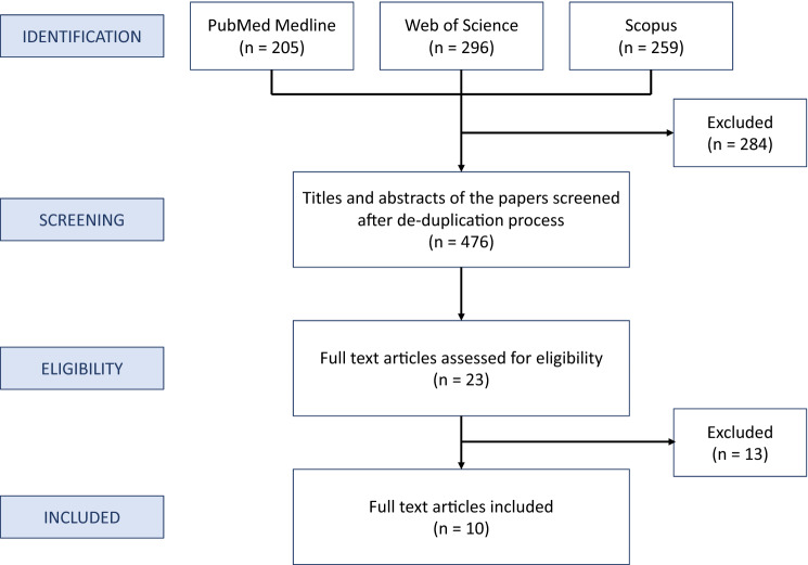
We conducted a systematic literature review on the use of [18F]FDG PET/MRI for staging/restaging rectal cancer patients with PubMed, Scopus, and Web of Science, based on the PRISMA criteria. Three authors screened all titles and abstracts and examined the full texts of all the identified relevant articles. Studies containing aggregated or duplicated data, review articles, case reports, editorials, and letters were excluded. Ten reports met the inclusion criteria. Four studies examined T staging and one focused on local recurrences after surgery; the reported sensitivity (94-100%), specificity (73-94%), and accuracy (92-100%) varied only slightly from one study to another. The sensitivity, specificity, and accuracy of [18F]FDG PET/MRI for N staging were 90-93%, 92-94%, and 42-92%. [18F]FDG PET/MRI detected malignant nodes better than MRI, resulting in treatment change. For M staging, [18F]FDG PET/MRI outperformed [18F]FDG PET/CT and CT in detecting liver metastases, whereas it performed worse for lung metastases. The results of this review suggest that [18F]FDG PET/MRI should be used for rectal cancer restaging after chemoradiotherapy and to select patients for rectum-sparing approaches thanks to its accuracy in T and N staging. For M staging, it should be associated at least with a chest CT scan to rule out lung metastases.
我们根据PRISMA标准,通过PubMed、Scopus和Web of Science对[18F]FDG PET/MRI用于直肠癌患者分期/再分期的情况进行了系统的文献综述。三位作者筛选了所有标题和摘要,并查阅了所有已识别的相关文章的全文。排除了包含汇总或重复数据的研究、综述文章、病例报告、社论和信件。十篇报告符合纳入标准。四项研究检查了T分期,一项研究关注术后局部复发;报告的敏感性(94%-100%)、特异性(73%-94%)和准确性(92%-100%)在不同研究之间仅有轻微差异。[18F]FDG PET/MRI对N分期的敏感性、特异性和准确性分别为90%-93%、92%-94%和42%-92%。[18F]FDG PET/MRI在检测恶性淋巴结方面优于MRI,从而导致治疗方案的改变。对于M分期,[18F]FDG PET/MRI在检测肝转移方面优于[18F]FDG PET/CT和CT,而在检测肺转移方面表现较差。本综述结果表明,由于[18F]FDG PET/MRI在T和N分期方面的准确性,应将其用于放化疗后的直肠癌再分期,并选择保留直肠的治疗方法的患者。对于M分期,它至少应与胸部CT扫描联合使用以排除肺转移。