Wu Yang, Liu Lu, Shen Xiaoyu, Liu Wenjing, Ma Rui
Medical Oncology Department of Thoracic Cancer (2), Cancer Hospital of China Medical University, Liaoning Cancer Hospital & Institute, Shenyang 110042, Liaoning, People's Republic of China.
Institute of Cancer Stem Cell, Dalian Medical University, Dalian 116044, Liaoning, People's Republic of China.
Cancer Manag Res. 2021 Jan 22;13:559-570. doi: 10.2147/CMAR.S281663. eCollection 2021.
Lung cancer is one of the most aggressive tumors with high incidence and mortality, which could be classified into lung squamous cell carcinoma (LUSC) and lung adenocarcinoma (LUAD). Overexpression of Plakophilin-2 (PKP2) has been reported in multiple malignancies. However, the expression and function mechanism of PKP2 in LUAD remain illusive.
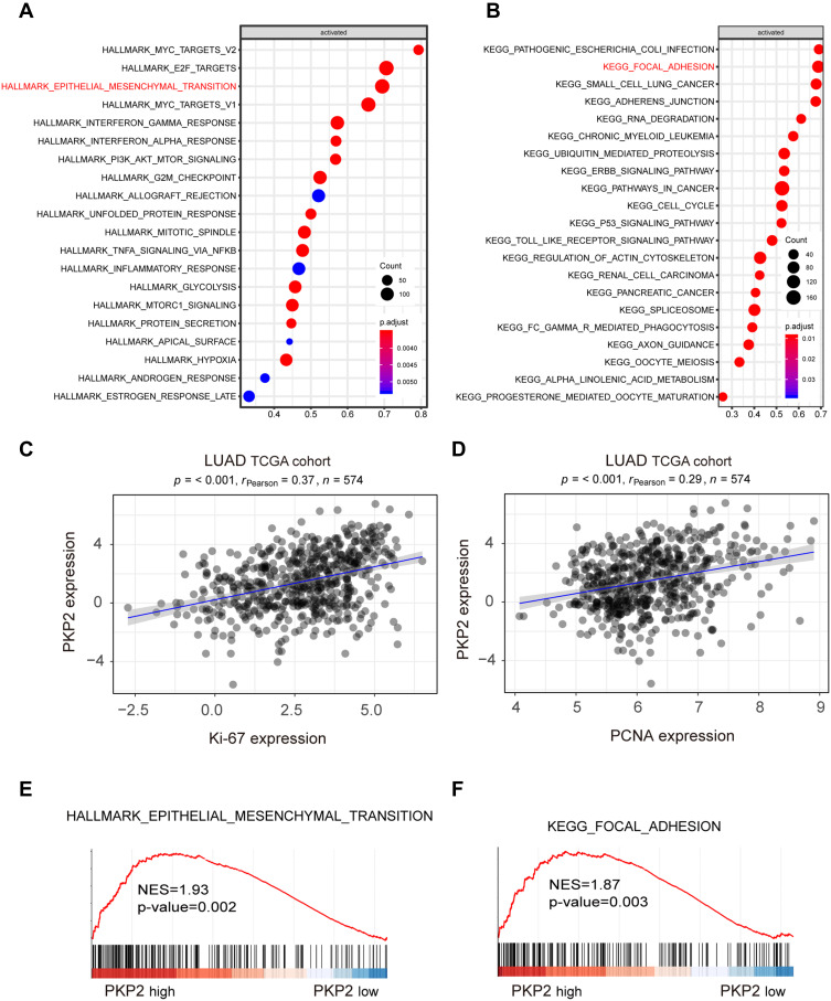
Real-time PCR (RT-PCR) was conducted to assess the expression of PKP2 in LUAD cells and tissues. An integrated analysis of PKP2 expression in The Cancer Genome Atlas (TCGA) was further performed. The effect of PKP2 on cell proliferation and invasion potential were then evaluated with loss-of-function assays in vitro. Xenograft nude mouse models were used to determine the role of PKP2 in LUAD tumorigenicity in vivo. Bioinformatics prediction, immunohistochemistry and Western blot were performed to examine whether PKP2 promoted LUAD development via enhancing focal adhesion and epithelial-mesenchymal transition.
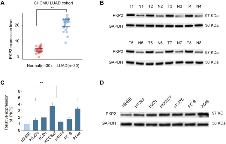
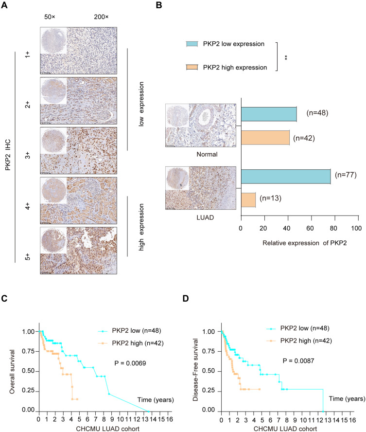
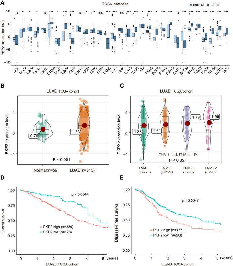
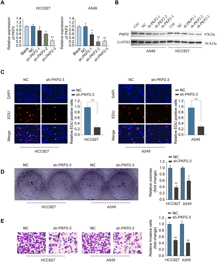
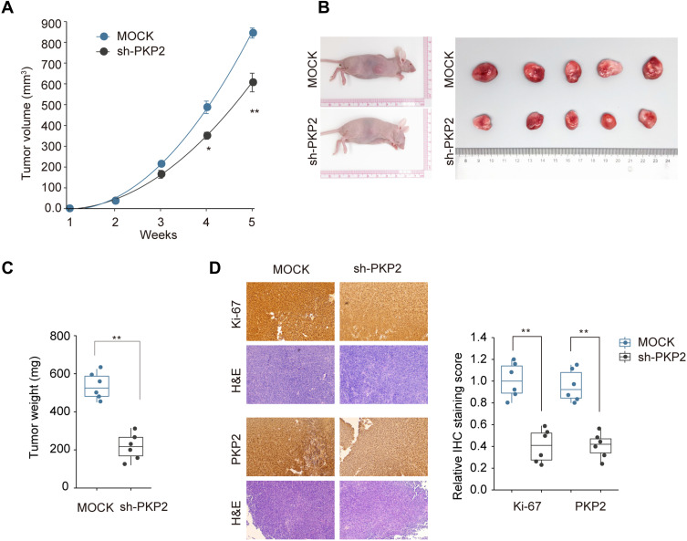
PKP2 expression was highly expressed in LUAD tissues compared with that in normal tissues and predicated poor prognosis of LUAD patients. TCGA LUAD cohort analysis also showed that high expression of PKP2 indicated unfavorable outcomes in LUAD patients. PKP2 expression was also upregulated in lung cancer cells. Functionally, knockdown of PKP2 suppressed lung cancer cell proliferation and invasion in vitro, while inhibited xenograft lung tumor development in vivo. Mechanistically, we demonstrated that high expression of PKP2 in LUAD was correlated with enhanced EMT and focal adhesion. Knockdown of PKP2 inhibited the expression of EMT-related Vimentin and N-cadherin and focal adhesion-associated expression of BMP4, ICAM1, and VCAM1 in xenograft tumors and lung cancer cells.
In summary, our findings indicate that PKP2 functions as an oncogene in LUAD, which could be utilized as a novel diagnostic and therapeutic marker for LUAD treatment.
肺癌是最具侵袭性的肿瘤之一,发病率和死亡率都很高,可分为肺鳞状细胞癌(LUSC)和肺腺癌(LUAD)。已有报道称,在多种恶性肿瘤中存在桥粒芯蛋白2(PKP2)的过表达。然而,PKP2在LUAD中的表达及功能机制仍不清楚。
采用实时荧光定量聚合酶链反应(RT-PCR)检测LUAD细胞和组织中PKP2的表达。进一步对癌症基因组图谱(TCGA)中PKP2的表达进行综合分析。然后通过体外功能缺失实验评估PKP2对细胞增殖和侵袭潜能的影响。利用异种移植裸鼠模型确定PKP2在LUAD体内致瘤性中的作用。进行生物信息学预测、免疫组织化学和蛋白质印迹分析,以检测PKP2是否通过增强黏着斑和上皮-间质转化促进LUAD的发展。
与正常组织相比,PKP2在LUAD组织中高表达,且提示LUAD患者预后不良。TCGA的LUAD队列分析也显示,PKP2的高表达表明LUAD患者预后不佳。PKP2在肺癌细胞中也上调。在功能上,敲低PKP2可抑制体外肺癌细胞的增殖和侵袭,同时抑制体内异种移植肺肿瘤的发展。机制上,我们证明了LUAD中PKP2的高表达与增强的上皮-间质转化和黏着斑相关。敲低PKP2可抑制异种移植肿瘤和肺癌细胞中上皮-间质转化相关的波形蛋白和N-钙黏蛋白的表达以及黏着斑相关的骨形态发生蛋白4、细胞间黏附分子1和血管细胞黏附分子1的表达。
总之,我们的研究结果表明PKP2在LUAD中作为一种癌基因发挥作用,可作为LUAD治疗的一种新的诊断和治疗标志物。