Liu Zi-Jun, Do Tiffany, Fong Hanson
Depts. Orthodontics & Oral Health Sciences, School of Dentistry.
Dept. Material Sciences and Engineering, College of Engineering, University of Washington, Seattle, WA, 98195, USA.
Heliyon. 2021 Jan 19;7(1):e05700. doi: 10.1016/j.heliyon.2020.e05700. eCollection 2021 Jan.
Obstructive sleep apnea (OSA) is associated with anatomical restrictions of pharyngeal airway, but the mechanism of airflow dynamics in OSA is largely unknown. This study utilized computational flow dynamics (CFD) to build a 3D model of the pharynx and to test the hypothesis that an increased restriction in the pharynx in OSA/obese minipigs leads to higher resistance, which in turn creates turbulence to induce temporary blockage of pharyngeal airway patency.
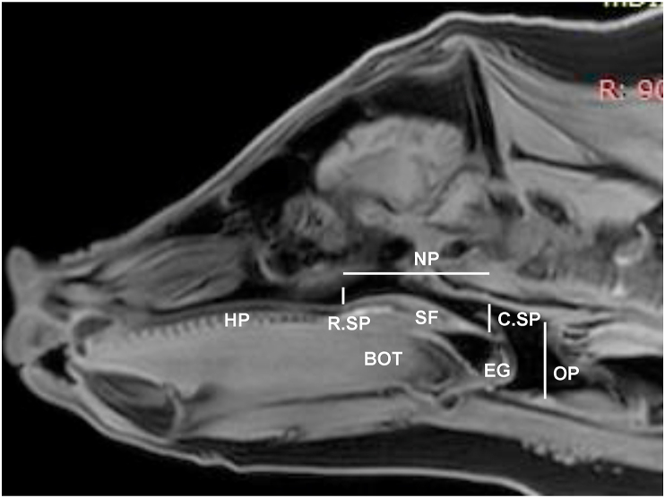
Of five 9-11-months-old Yucatan minipigs, 3 were non-obese (BMI<35) and two obese (BMI>51). After natural sleep monitoring using BioRadio system, pigs were sedated to collect MRI images and airflow parameters. The MRI images were processed to create 3D configurations of pharynx. These 3D configurations were meshed to create finite element models (FEM) of CFD. The obtained airflow parameters were input into the configurations to identify turbulent airflow and its location.
Heavy snoring and multiple >5s hypopnea/apnea episodes (AHI = 32-35) were identified in both obese minipigs during sleep. Compared to the non-obese/non-OSA controls, obese/OSA minipigs showed much lower respiratory tidal volumes and inspiratory airflow speed. FEM simulation found that turbulence was not present in the pharynx in either model. However, a 25% increase of airflow velocity was observed at the narrowest part of the nasal pharynx in the obese/OSA minipig model.
Despite the narrower pharyngeal airway and the higher velocity of airflow, FEM simulation indicated that turbulence was not produced in the obese/OSA minipigs.
阻塞性睡眠呼吸暂停(OSA)与咽气道的解剖学限制有关,但OSA中气流动力学的机制在很大程度上尚不清楚。本研究利用计算流体动力学(CFD)构建咽部的三维模型,并检验以下假设:OSA/肥胖小型猪咽部限制增加会导致更高的阻力,进而产生湍流,导致咽部气道通畅性暂时受阻。
在5只9 - 11月龄的尤卡坦小型猪中,3只为非肥胖(BMI<35),2只为肥胖(BMI>51)。使用生物无线电系统进行自然睡眠监测后,对猪进行镇静以收集MRI图像和气流参数。对MRI图像进行处理以创建咽部的三维结构。将这些三维结构网格化以创建CFD的有限元模型(FEM)。将获得的气流参数输入到这些结构中,以识别湍流气流及其位置。
在睡眠期间,两只肥胖小型猪均出现严重打鼾和多次>5秒的呼吸暂停/低通气发作(AHI = 32 - 35)。与非肥胖/非OSA对照组相比,肥胖/OSA小型猪的呼吸潮气量和吸气气流速度要低得多。有限元模型模拟发现,两个模型的咽部均未出现湍流。然而,在肥胖/OSA小型猪模型的鼻咽最窄处观察到气流速度增加了25%。
尽管咽部气道较窄且气流速度较高,但有限元模型模拟表明,肥胖/OSA小型猪并未产生湍流。