Kuang Huihui, Schneiderman Zachary, Shabana Ahmed M, Russo Gabriella C, Guo Jun, Wirtz Denis, Kokkoli Efrosini
Institute for NanoBioTechnology Johns Hopkins University Baltimore Maryland USA.
Department of Chemical and Biomolecular Engineering Johns Hopkins University Baltimore Maryland USA.
Bioeng Transl Med. 2020 Nov 10;6(1):e10194. doi: 10.1002/btm2.10194. eCollection 2021 Jan.
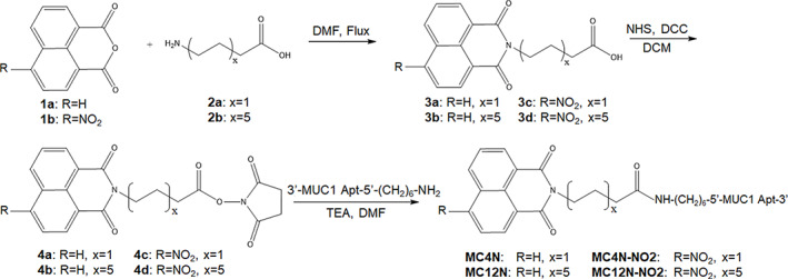
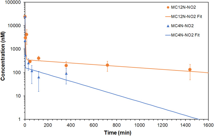
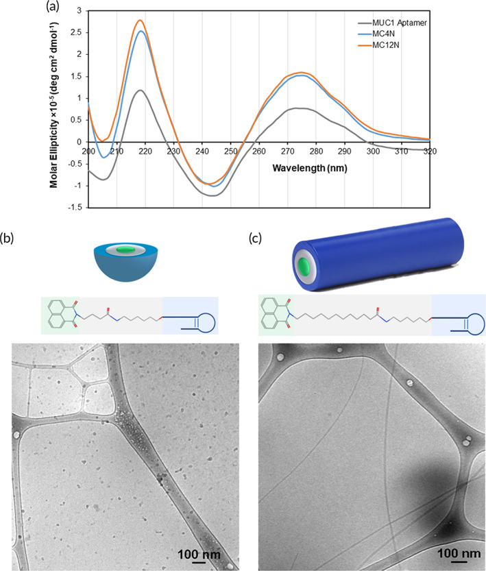
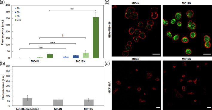
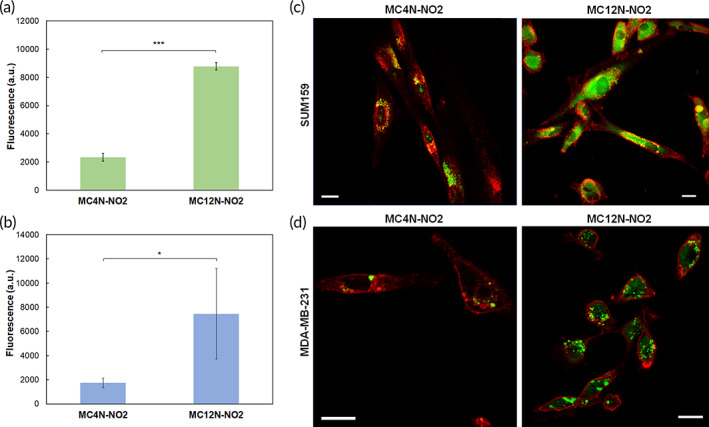
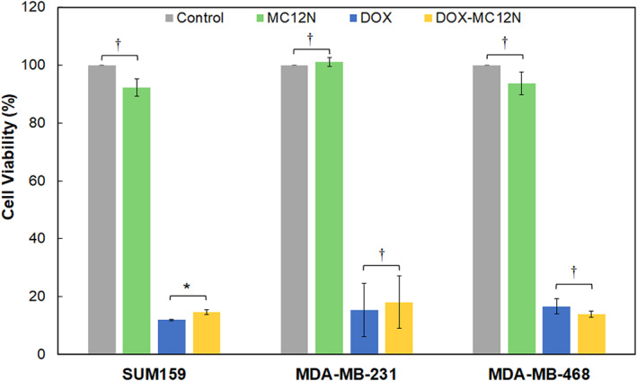
Despite decades of research, there are few targeted treatment options available for triple negative breast cancer (TNBC), leaving chemotherapy, and radiation treatment regimes with poor response and high toxicity. Herein aptamer-amphiphiles were synthesized which selectively bind to the mucin-1 (MUC1) glycoprotein that is overexpressed in TNBC cells. These amphiphiles have a fluorescent tail (1,8-naphthalimide or 4-nitro-1,8-naphthalimide) which enables self-assembly of the amphiphiles and allows for easy visualization without the requirement for further conjugation of a fluorophore. Interestingly, the length of the alkyl spacer (C or C) between the aptamer and tail was shown to influence the morphology of the self-assembled structure, and thus its ability to internalize into the TNBC cells. While both the MUC1 aptamer-C-napthalimide spherical micelles and the MUC1 aptamer-C-napthalimide long cylindrical micelles showed internalization into MDA-MB-468 TNBC cells but not the noncancerous MCF-10A breast cells, the cylindrical micelles showed greatly enhanced internalization into the MDA-MB-468 cells. Similar patterns of enhanced binding and internalization were observed between the MUC1 aptamer-C-napthalimide cylindrical micelles and SUM159 and MDA-MB-231 TNBC cells. The MUC1 aptamer cylindrical micelles were not toxic to the cells, and when used to deliver doxorubicin to the TNBC cells, were shown to be as cytotoxic as free doxorubicin. Moreover, a pharmacokinetic study in mice showed a prolonged systemic circulation time of the MUC1 aptamer cylindrical micelles. There was a 4.6-fold increase in the elimination half-life of the aptamer cylindrical micelles, and their clearance decreased 10-fold compared to the MUC1 aptamer spherical micelles. Thus, the MUC1 aptamer-C-napthalimide nanofibers represent a promising vehicle that could be used for easy visualization and targeted delivery of therapeutic loads to TNBC cells.
尽管经过了数十年的研究,但三阴性乳腺癌(TNBC)几乎没有靶向治疗方案,化疗和放射治疗方案的反应不佳且毒性高。在此合成了适体两亲物,其选择性结合在TNBC细胞中过表达的粘蛋白-1(MUC1)糖蛋白。这些两亲物具有荧光尾巴(1,8-萘二甲酰亚胺或4-硝基-1,8-萘二甲酰亚胺),这使得两亲物能够自组装,并无需进一步连接荧光团即可轻松可视化。有趣的是,适体与尾巴之间的烷基间隔基(C或C)的长度显示会影响自组装结构的形态,从而影响其内化进入TNBC细胞的能力。虽然MUC1适体-C-萘二甲酰亚胺球形胶束和MUC1适体-C-萘二甲酰亚胺长圆柱形胶束均显示内化进入MDA-MB-468 TNBC细胞,但未进入非癌性MCF-10A乳腺细胞,圆柱形胶束显示进入MDA-MB-468细胞的内化作用大大增强。在MUC1适体-C-萘二甲酰亚胺圆柱形胶束与SUM159和MDA-MB-231 TNBC细胞之间观察到了类似的增强结合和内化模式。MUC1适体圆柱形胶束对细胞无毒,并且当用于将阿霉素递送至TNBC细胞时,显示出与游离阿霉素一样的细胞毒性。此外,在小鼠中进行的药代动力学研究表明,MUC1适体圆柱形胶束的全身循环时间延长。适体圆柱形胶束的消除半衰期增加了4.6倍,与MUC1适体球形胶束相比,其清除率降低了10倍。因此,MUC1适体-C-萘二甲酰亚胺纳米纤维代表了一种有前途的载体,可用于轻松可视化并将治疗载荷靶向递送至TNBC细胞。