• 文献检索
  • 文档翻译
  • 深度研究
  • 学术资讯
  • Suppr Zotero 插件Zotero 插件
  • 邀请有礼
  • 套餐&价格
  • 历史记录
应用&插件
Suppr Zotero 插件Zotero 插件浏览器插件Mac 客户端Windows 客户端微信小程序
定价
高级版会员购买积分包购买API积分包
服务
文献检索文档翻译深度研究API 文档MCP 服务
关于我们
关于 Suppr公司介绍联系我们用户协议隐私条款
关注我们

Suppr 超能文献

核心技术专利:CN118964589B侵权必究
粤ICP备2023148730 号-1Suppr @ 2026

文献检索

告别复杂PubMed语法,用中文像聊天一样搜索,搜遍4000万医学文献。AI智能推荐,让科研检索更轻松。

立即免费搜索

文件翻译

保留排版,准确专业,支持PDF/Word/PPT等文件格式,支持 12+语言互译。

免费翻译文档

深度研究

AI帮你快速写综述,25分钟生成高质量综述,智能提取关键信息,辅助科研写作。

立即免费体验

SARS-CoV-2 通过 UNC93B 和 IRAK4 诱导人类浆细胞前树突状细胞多样化。

SARS-CoV-2 induces human plasmacytoid predendritic cell diversification via UNC93B and IRAK4.

机构信息

Université de Paris, Institut de Recherche Saint-Louis, Institut National de la Santé et de la Recherche Médicale U976, Hôpital Saint-Louis, Paris, France.

Université de Paris, Institut de Recherche Saint-Louis, Institut National de la Santé et de la Recherche Médicale U944, Centre National de la Recherche Scientifique 7212, Hôpital Saint-Louis, Paris, France.

出版信息

J Exp Med. 2021 Apr 5;218(4). doi: 10.1084/jem.20201387.

DOI:10.1084/jem.20201387
PMID:33533916
原文链接:https://pmc.ncbi.nlm.nih.gov/articles/PMC7849819/
Abstract

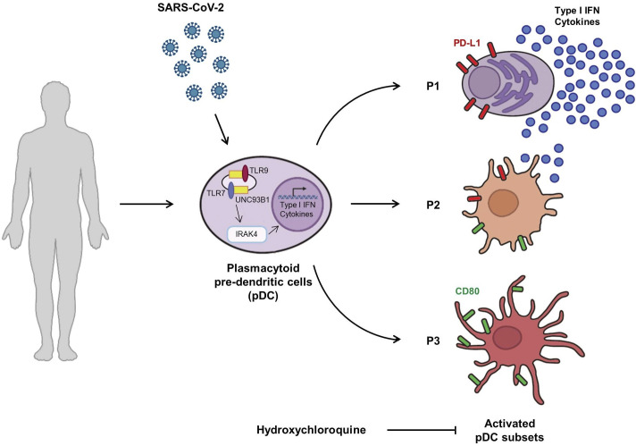
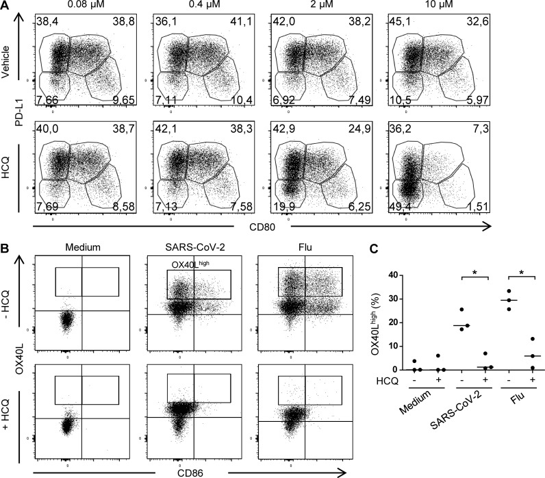
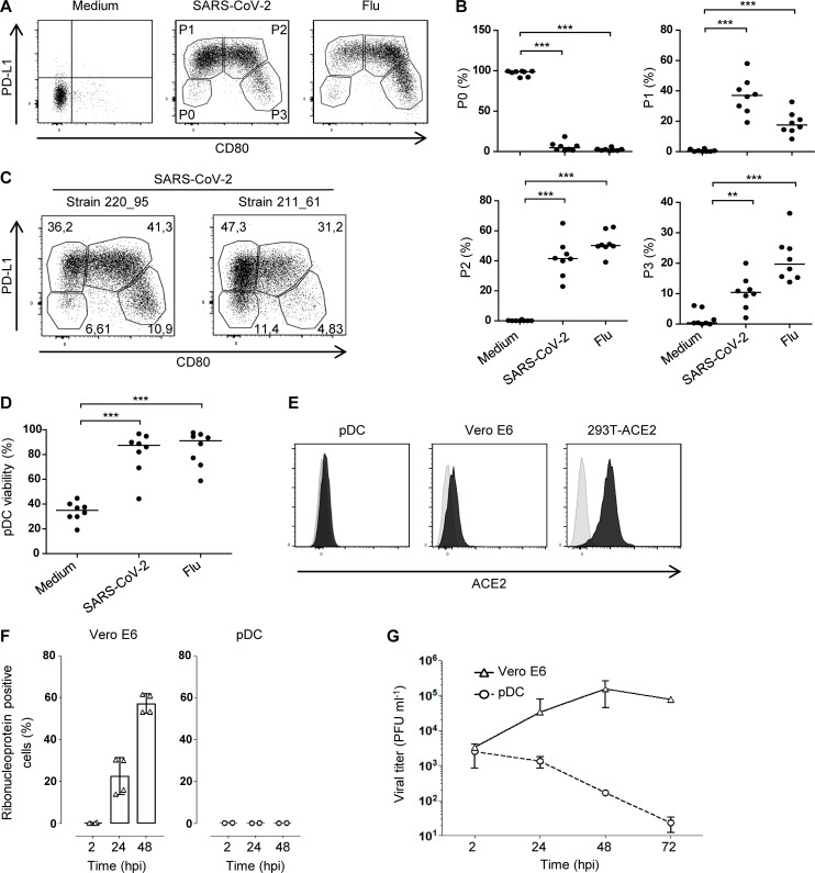
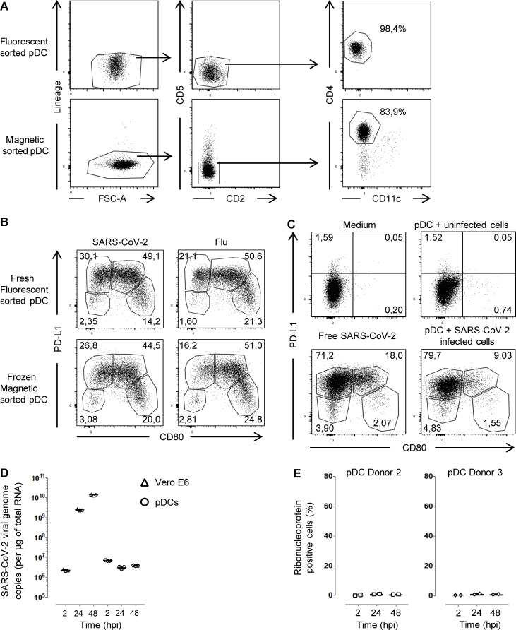
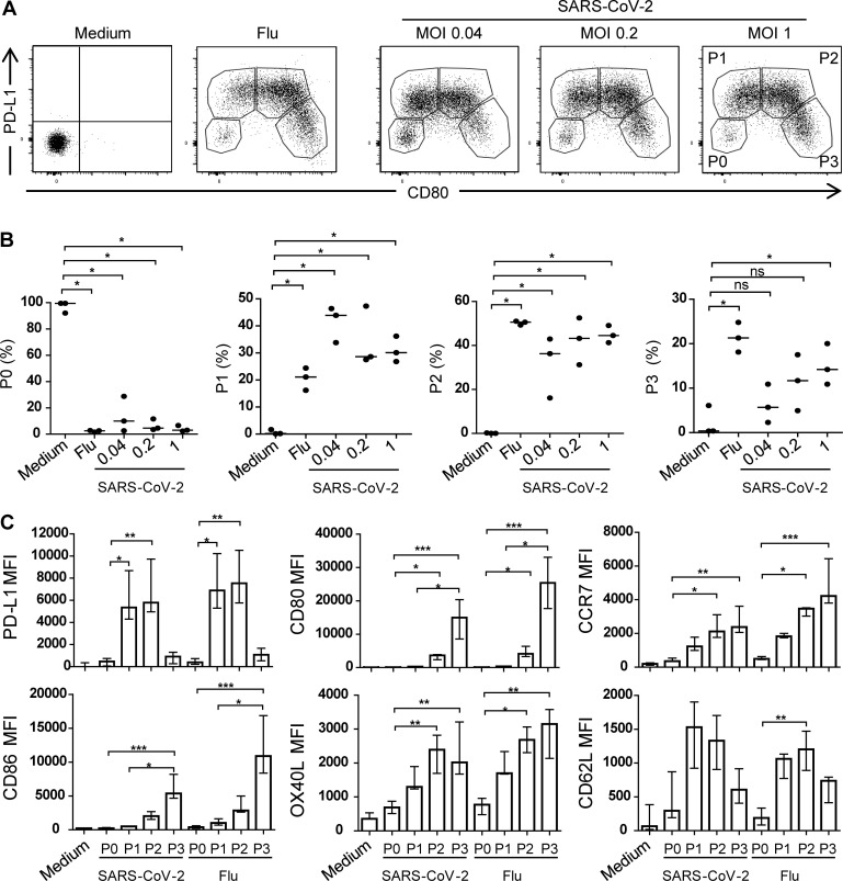
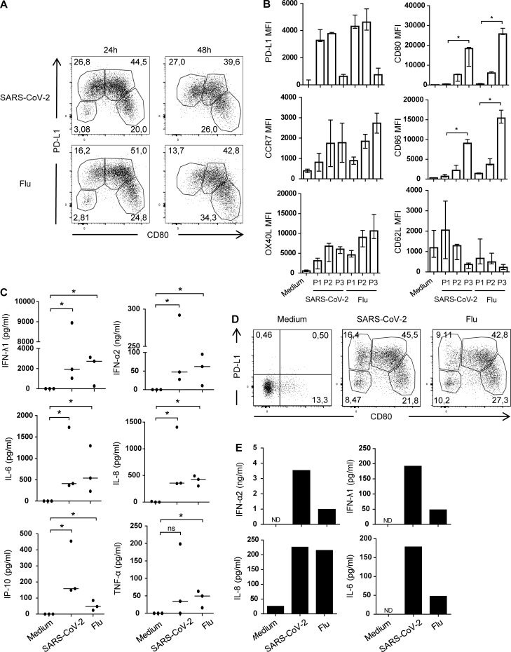
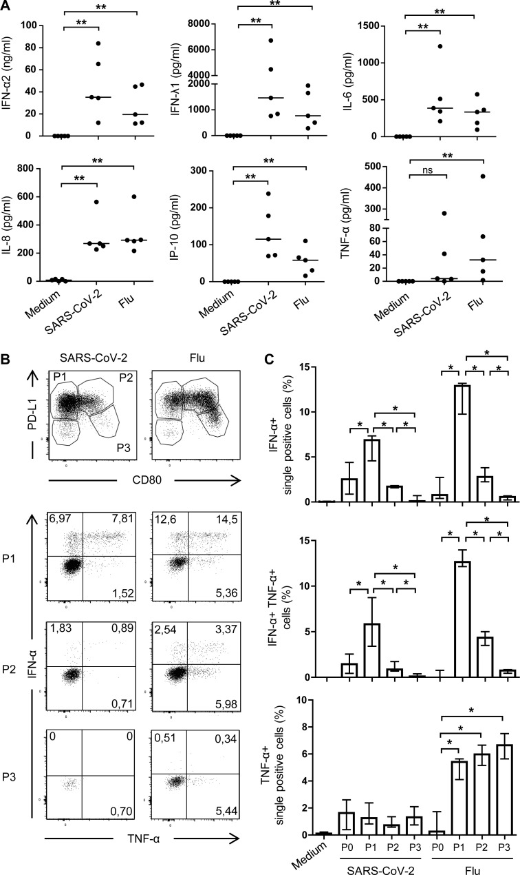
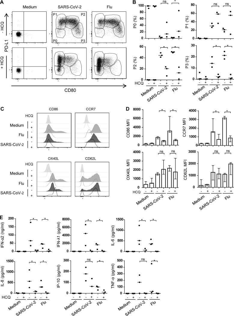
Several studies have analyzed antiviral immune pathways in late-stage severe COVID-19. However, the initial steps of SARS-CoV-2 antiviral immunity are poorly understood. Here we have isolated primary SARS-CoV-2 viral strains and studied their interaction with human plasmacytoid predendritic cells (pDCs), a key player in antiviral immunity. We show that pDCs are not productively infected by SARS-CoV-2. However, they efficiently diversified into activated P1-, P2-, and P3-pDC effector subsets in response to viral stimulation. They expressed CD80, CD86, CCR7, and OX40 ligand at levels similar to influenza virus-induced activation. They rapidly produced high levels of interferon-α, interferon-λ1, IL-6, IP-10, and IL-8. All major aspects of SARS-CoV-2-induced pDC activation were inhibited by hydroxychloroquine. Mechanistically, SARS-CoV-2-induced pDC activation critically depended on IRAK4 and UNC93B1, as established using pDC from genetically deficient patients. Overall, our data indicate that human pDC are efficiently activated by SARS-CoV-2 particles and may thus contribute to type I IFN-dependent immunity against SARS-CoV-2 infection.

摘要

几项研究分析了晚期严重 COVID-19 中的抗病毒免疫途径。然而,人们对 SARS-CoV-2 抗病毒免疫的初始步骤知之甚少。在这里,我们分离了原代 SARS-CoV-2 病毒株,并研究了它们与人浆细胞样树突状细胞(pDC)的相互作用,pDC 是抗病毒免疫的关键细胞。我们发现 pDC 不能被 SARS-CoV-2 有效感染。然而,它们在受到病毒刺激时能够有效地多样化为激活的 P1、P2 和 P3-pDC 效应亚群。它们表达 CD80、CD86、CCR7 和 OX40 配体的水平与流感病毒诱导的激活相似。它们迅速产生高水平的干扰素-α、干扰素-λ1、IL-6、IP-10 和 IL-8。氯喹可抑制 SARS-CoV-2 诱导的 pDC 活化的所有主要方面。从机制上讲,SARS-CoV-2 诱导的 pDC 活化严重依赖于 IRAK4 和 UNC93B1,这是使用基因缺陷患者的 pDC 确定的。总体而言,我们的数据表明,人类 pDC 可被 SARS-CoV-2 颗粒有效激活,因此可能有助于依赖 I 型 IFN 的 SARS-CoV-2 感染免疫。

https://cdn.ncbi.nlm.nih.gov/pmc/blobs/151e/7849819/24576229f7eb/JEM_20201387_FigS3.jpg
https://cdn.ncbi.nlm.nih.gov/pmc/blobs/151e/7849819/4d78af1b9d0e/JEM_20201387_GA.jpg
https://cdn.ncbi.nlm.nih.gov/pmc/blobs/151e/7849819/3721cf89cccd/JEM_20201387_Fig1.jpg
https://cdn.ncbi.nlm.nih.gov/pmc/blobs/151e/7849819/70ffb4fdc0be/JEM_20201387_FigS1.jpg
https://cdn.ncbi.nlm.nih.gov/pmc/blobs/151e/7849819/68954334cadf/JEM_20201387_Fig2.jpg
https://cdn.ncbi.nlm.nih.gov/pmc/blobs/151e/7849819/087b3f3c1319/JEM_20201387_FigS2.jpg
https://cdn.ncbi.nlm.nih.gov/pmc/blobs/151e/7849819/1dcbf28a9967/JEM_20201387_Fig3.jpg
https://cdn.ncbi.nlm.nih.gov/pmc/blobs/151e/7849819/5dfbe69e934d/JEM_20201387_Fig4.jpg
https://cdn.ncbi.nlm.nih.gov/pmc/blobs/151e/7849819/24576229f7eb/JEM_20201387_FigS3.jpg

相似文献

1
SARS-CoV-2 induces human plasmacytoid predendritic cell diversification via UNC93B and IRAK4.SARS-CoV-2 通过 UNC93B 和 IRAK4 诱导人类浆细胞前树突状细胞多样化。
J Exp Med. 2021 Apr 5;218(4). doi: 10.1084/jem.20201387.
2
SARS-CoV-2 induces human plasmacytoid pre-dendritic cell diversification via UNC93B and IRAK4.严重急性呼吸综合征冠状病毒2通过UNC93B和白细胞介素-1受体相关激酶4诱导人浆细胞样前树突状细胞分化。
bioRxiv. 2021 Jan 8:2020.07.10.197343. doi: 10.1101/2020.07.10.197343.
3
Macrophages and Dendritic Cells Are Not the Major Source of Pro-Inflammatory Cytokines Upon SARS-CoV-2 Infection.巨噬细胞和树突状细胞不是新冠病毒感染后促炎细胞因子的主要来源。
Front Immunol. 2021 May 26;12:647824. doi: 10.3389/fimmu.2021.647824. eCollection 2021.
4
Type I Interferon Induction and Exhaustion during Viral Infection: Plasmacytoid Dendritic Cells and Emerging COVID-19 Findings.病毒感染期间的 I 型干扰素诱导和耗竭:浆细胞样树突状细胞和新兴的 COVID-19 研究结果。
Viruses. 2021 Sep 15;13(9):1839. doi: 10.3390/v13091839.
5
Severe COVID-19 patients have impaired plasmacytoid dendritic cell-mediated control of SARS-CoV-2.重症 COVID-19 患者的浆细胞样树突状细胞介导的 SARS-CoV-2 控制受损。
Nat Commun. 2023 Feb 8;14(1):694. doi: 10.1038/s41467-023-36140-9.
6
Differential plasmacytoid dendritic cell phenotype and type I Interferon response in asymptomatic and severe COVID-19 infection.无症状和重症 COVID-19 感染中的浆细胞样树突状细胞表型和 I 型干扰素反应的差异。
PLoS Pathog. 2021 Sep 2;17(9):e1009878. doi: 10.1371/journal.ppat.1009878. eCollection 2021 Sep.
7
TLR2 and TLR7 mediate distinct immunopathological and antiviral plasmacytoid dendritic cell responses to SARS-CoV-2 infection.Toll样受体2(TLR2)和Toll样受体7(TLR7)介导浆细胞样树突状细胞对严重急性呼吸综合征冠状病毒2(SARS-CoV-2)感染产生不同的免疫病理反应和抗病毒反应。
EMBO J. 2022 May 16;41(10):e109622. doi: 10.15252/embj.2021109622. Epub 2022 Mar 1.
8
Macrophage phagocytosis of SARS-CoV-2-infected cells mediates potent plasmacytoid dendritic cell activation.巨噬细胞吞噬 SARS-CoV-2 感染的细胞可介导强烈的浆细胞样树突状细胞激活。
Cell Mol Immunol. 2023 Jul;20(7):835-849. doi: 10.1038/s41423-023-01039-4. Epub 2023 May 30.
9
Age-Related Expression of IFN-λ1 IFN-I and Beta-Defensins in the Nasopharynx of SARS-CoV-2-Infected Individuals.年龄相关性 IFN-λ1、IFN-I 和 β 防御素在 SARS-CoV-2 感染个体鼻咽部的表达。
Front Immunol. 2021 Nov 10;12:750279. doi: 10.3389/fimmu.2021.750279. eCollection 2021.
10
Antiviral Activity of Type I, II, and III Interferons Counterbalances ACE2 Inducibility and Restricts SARS-CoV-2.I型、II型和III型干扰素的抗病毒活性可抵消ACE2的诱导性并限制新型冠状病毒。
mBio. 2020 Sep 10;11(5):e01928-20. doi: 10.1128/mBio.01928-20.

引用本文的文献

1
In Vitro Investigation of Statin Effects on Genes Associated with Severe COVID-19 in Cancerous and Non-Cancerous Cells.他汀类药物对癌细胞和非癌细胞中与重症 COVID-19 相关基因影响的体外研究
Biomedicines. 2025 Jul 14;13(7):1714. doi: 10.3390/biomedicines13071714.
2
Modulation of IRAK4 as a Therapeutic Strategy Against Monosodium Urate- and Xanthine-Induced Inflammation in Macrophages and HepG2 Cells.调节白细胞介素-1受体相关激酶4作为对抗尿酸钠和黄嘌呤诱导的巨噬细胞和肝癌细胞系炎症的治疗策略
Res Sq. 2025 Jul 16:rs.3.rs-6908346. doi: 10.21203/rs.3.rs-6908346/v1.
3
Enhanced TLR7-dependent production of type I interferon by pDCs underlies pandemic chilblains.

本文引用的文献

1
Life-Threatening COVID-19: Defective Interferons Unleash Excessive Inflammation.危及生命的 COVID-19:缺陷型干扰素引发过度炎症。
Med. 2020 Dec 18;1(1):14-20. doi: 10.1016/j.medj.2020.12.001.
2
COVID-19: Discovery, diagnostics and drug development.新型冠状病毒肺炎:发现、诊断和药物研发。
J Hepatol. 2021 Jan;74(1):168-184. doi: 10.1016/j.jhep.2020.09.031. Epub 2020 Oct 8.
3
Autoantibodies against type I IFNs in patients with life-threatening COVID-19.COVID-19 危重症患者体内针对 I 型干扰素的自身抗体。
浆细胞样树突状细胞(pDCs)中由Toll样受体7(TLR7)依赖性增强的I型干扰素产生是大流行冻疮的基础。
J Exp Med. 2025 Jul 7;222(7). doi: 10.1084/jem.20231467. Epub 2025 Apr 14.
4
Characterization of TLR9 responsiveness in cell subsets derived from pDC differentiation of hematopoietic stem and progenitor cells.造血干细胞和祖细胞浆细胞样树突状细胞分化来源的细胞亚群中Toll样受体9反应性的特征分析
Front Immunol. 2025 Mar 27;16:1550397. doi: 10.3389/fimmu.2025.1550397. eCollection 2025.
5
Toll-like receptor 7 (TLR7)-mediated antiviral response protects mice from lethal SARS-CoV-2 infection.Toll样受体7(TLR7)介导的抗病毒反应可保护小鼠免受致命的严重急性呼吸综合征冠状病毒2(SARS-CoV-2)感染。
J Virol. 2025 May 20;99(5):e0166824. doi: 10.1128/jvi.01668-24. Epub 2025 Mar 31.
6
DOCK2 Deficiency and GATA2 Haploinsufficiency Can Underlie Critical Coronavirus Disease 2019 (COVID-19) Pneumonia.DOCK2缺陷和GATA2单倍体不足可能是重症2019冠状病毒病(COVID-19)肺炎的潜在病因。
J Clin Immunol. 2025 Mar 28;45(1):85. doi: 10.1007/s10875-025-01877-z.
7
Role of innate immunity in SARS-CoV-2 infection.固有免疫在2019冠状病毒病感染中的作用。
Biosaf Health. 2023 Sep 9;5(5):280-288. doi: 10.1016/j.bsheal.2023.08.005. eCollection 2023 Oct.
8
SARS-CoV-2 S, M, and E Structural Glycoproteins Differentially Modulate Endoplasmic Reticulum Stress Responses.严重急性呼吸综合征冠状病毒2型的刺突蛋白、膜蛋白和包膜蛋白对内质网应激反应有不同的调节作用。
Int J Mol Sci. 2025 Jan 26;26(3):1047. doi: 10.3390/ijms26031047.
9
Recent Advances in Our Understanding of Human Inflammatory Dendritic Cells in Human Immunodeficiency Virus Infection.我们对人类免疫缺陷病毒感染中人类炎性树突状细胞理解的最新进展
Viruses. 2025 Jan 14;17(1):105. doi: 10.3390/v17010105.
10
Sensing of SARS-CoV-2-infected cells by plasmacytoid dendritic cells is modulated via an interplay between CD54/ICAM-1 and CD11a/LFA-1 α integrin.浆细胞样树突状细胞对感染严重急性呼吸综合征冠状病毒2(SARS-CoV-2)的细胞的感知通过CD54/细胞间黏附分子-1(ICAM-1)和CD11a/淋巴细胞功能相关抗原-1(LFA-1)α整合素之间的相互作用来调节。
J Virol. 2025 Feb 25;99(2):e0123524. doi: 10.1128/jvi.01235-24. Epub 2025 Jan 13.
Science. 2020 Oct 23;370(6515). doi: 10.1126/science.abd4585. Epub 2020 Sep 24.
4
Inborn errors of type I IFN immunity in patients with life-threatening COVID-19.COVID-19 危重症患者的 I 型 IFN 免疫先天缺陷。
Science. 2020 Oct 23;370(6515). doi: 10.1126/science.abd4570. Epub 2020 Sep 24.
5
Presence of Genetic Variants Among Young Men With Severe COVID-19.年轻男性严重 COVID-19 患者中存在遗传变异。
JAMA. 2020 Aug 18;324(7):663-673. doi: 10.1001/jama.2020.13719.
6
COVID-19: Emergence, Spread, Possible Treatments, and Global Burden.新型冠状病毒肺炎(COVID-19):起源、传播、可能的治疗方法及全球负担。
Front Public Health. 2020 May 28;8:216. doi: 10.3389/fpubh.2020.00216. eCollection 2020.
7
The kinase IRAK4 promotes endosomal TLR and immune complex signaling in B cells and plasmacytoid dendritic cells.激酶 IRAK4 促进了 B 细胞和浆细胞样树突状细胞中内体 TLR 和免疫复合物信号转导。
Sci Signal. 2020 Jun 2;13(634):eaaz1053. doi: 10.1126/scisignal.aaz1053.
8
An Updated Systematic Review of the Therapeutic Role of Hydroxychloroquine in Coronavirus Disease-19 (COVID-19).羟氯喹在新型冠状病毒肺炎(COVID-19)治疗作用的更新系统评价。
Clin Drug Investig. 2020 Jul;40(7):591-601. doi: 10.1007/s40261-020-00927-1.
9
Dysregulation of type I interferon responses in COVID-19.COVID-19 中 I 型干扰素反应失调。
Nat Rev Immunol. 2020 Jul;20(7):397-398. doi: 10.1038/s41577-020-0346-x. Epub 2020 May 26.
10
The hallmarks of COVID-19 disease.新型冠状病毒疾病的特征。
PLoS Pathog. 2020 May 22;16(5):e1008536. doi: 10.1371/journal.ppat.1008536. eCollection 2020 May.