Guangxi Medical University, Nanning, 530021, People's Republic of China.
Department of Oral and Maxillofacial Surgery, Hospital of Stomatology, Guangxi Medical University, Nanning, 530021, People's Republic of China.
Stem Cell Res Ther. 2021 Feb 3;12(1):101. doi: 10.1186/s13287-021-02150-x.
Distraction osteogenesis (DO) is a highly efficacious form of reconstructive bone regeneration, but its clinical utility is limited by the prolonged period required for bone consolidation to occur. Understanding the mechanistic basis for DO and shortening this consolidation phase thus represent promising approaches to improving the clinical utility of this procedure.
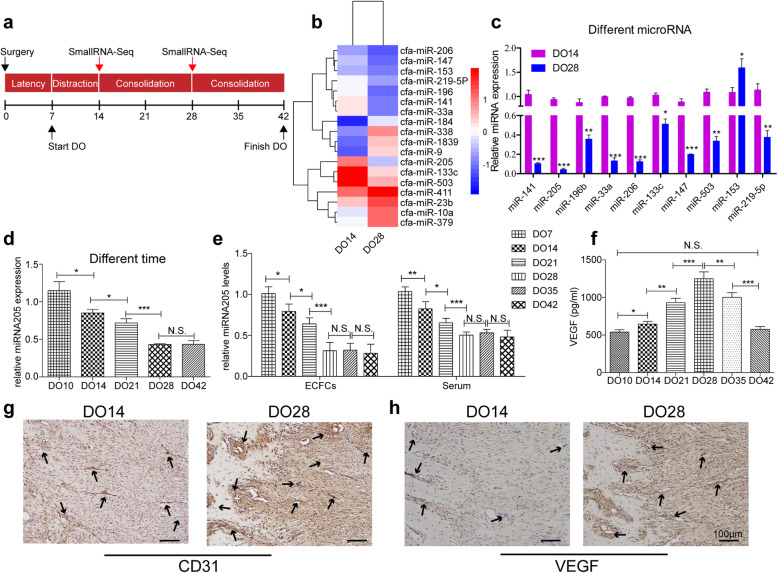
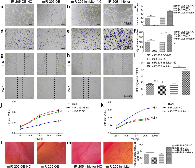
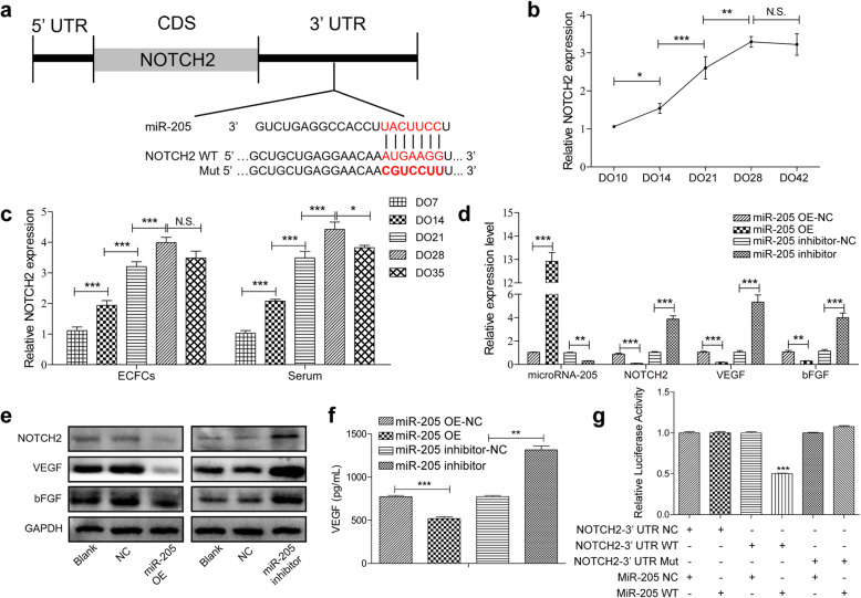
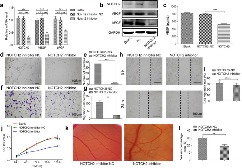
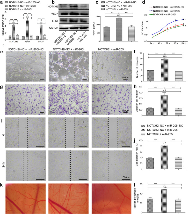
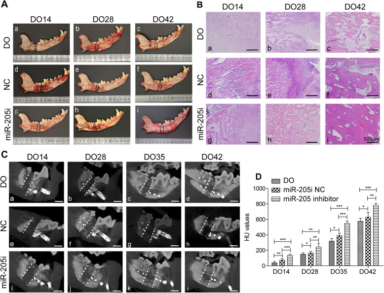
A mandibular DO (MDO) canine model was established, after which small RNA sequencing was performed to identify relevant molecular targets genes. Putative miRNA target genes were identified through bioinformatics and confirmed through qPCR, Western blotting, and dual-luciferase reporter assays. Peripheral blood samples were collected to isolate serum and endothelial colony-forming cells (ECFCs) in order to measure miR-205, NOTCH2, and angiogenic cytokines expression levels. Lentiviral constructs were then used to inhibit or overexpress miR-205 and NOTCH2 in isolated ECFCs, after which the angiogenic activity of these cells was evaluated in migration, wound healing, proliferation, tube formation, and chick chorioallantoic membrane (CAM) assay. Autologous ECFCs transfected to knockdown miR-205 and were injected directly into the distraction callus. On days 14, 28, 35 and 42 after surgery, bone density was evaluated via CBCT, and callus samples were collected and evaluated via histological staining to analyze bone regeneration and remodeling.
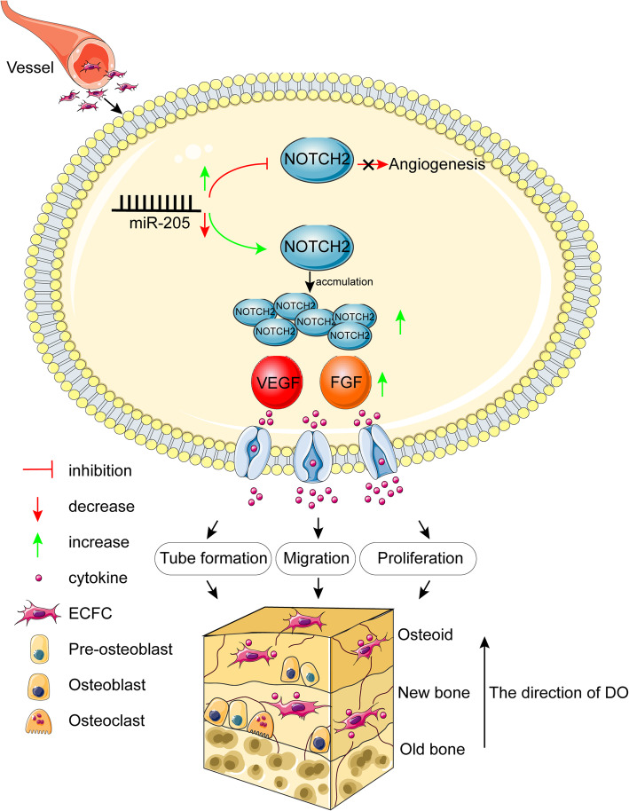
MiR-205 was identified as being one of the miRNAs that was most significantly downregulated in MDO callus samples. Downregulation of miR-205 was also observed in DO-ECFCs and serum of animals undergoing MDO. Inhibiting miR-205 markedly enhanced angiogenesis, whereas overexpressing miR-205 had the opposite effect in vitro. Importantly, NOTCH2, which is a unique regulator in bone angiogenesis, was identified as a miR-205 target gene. Consistent with this regulatory relationship, knocking down NOTCH2 suppressed angiogenesis, and transduction with a miR-205 inhibitor lentivirus was sufficient to rescue angiogenic activity. When ECFCs in which miR-205 had been inhibited were transplanted into the MDO callus, this significantly bolstered osteogenesis, and remodeling in vivo.
MiR-205 is a significant regulator of the MDO process, and inhibiting this miRNA can accelerate MDO-related mineralization. Overall, these results offer new insights into the mechanistic basis for this procedure, highlighting potential targets for therapeutic clinical intervention.
牵张成骨术(DO)是一种高效的重建性骨再生形式,但由于骨整合所需的时间较长,其临床应用受到限制。了解 DO 的机制基础并缩短这个整合阶段,因此代表了改善该手术临床应用的有前途的方法。
建立了下颌骨 DO(MDO)犬模型,然后进行小 RNA 测序以鉴定相关的分子靶标基因。通过生物信息学和 qPCR、Western blot 和双荧光素酶报告基因分析鉴定推定的 miRNA 靶基因。收集外周血样本以分离血清和内皮祖细胞(ECFCs),以测量 miR-205、NOTCH2 和血管生成细胞因子的表达水平。然后使用慢病毒构建体抑制或过表达分离的 ECFCs 中的 miR-205 和 NOTCH2,之后在迁移、划痕愈合、增殖、管形成和鸡胚绒毛尿囊膜(CAM)分析中评估这些细胞的血管生成活性。将转染了 miR-205 敲低的自体 ECFCs 直接注射到牵张性骨痂中。在手术第 14、28、35 和 42 天,通过 CBCT 评估骨密度,并采集骨痂样本进行组织学染色,以分析骨再生和重塑。
miR-205 被鉴定为 MDO 骨痂样本中下调最显著的 miRNA 之一。在 DO-ECFCs 和经历 MDO 的动物的血清中也观察到 miR-205 的下调。抑制 miR-205 可显著增强血管生成,而体外过表达 miR-205 则有相反的效果。重要的是,NOTCH2,一种骨血管生成的独特调节剂,被鉴定为 miR-205 的靶基因。与这种调节关系一致,敲低 NOTCH2 抑制血管生成,转导 miR-205 抑制剂慢病毒足以恢复血管生成活性。当将抑制 miR-205 的 ECFCs 移植到 MDO 骨痂中时,这显著增强了体内成骨和重塑。
miR-205 是 MDO 过程的重要调节剂,抑制这种 miRNA 可以加速 MDO 相关的矿化。总的来说,这些结果为该手术的机制基础提供了新的见解,突出了治疗性临床干预的潜在靶点。