• 文献检索
  • 文档翻译
  • 深度研究
  • 学术资讯
  • Suppr Zotero 插件Zotero 插件
  • 邀请有礼
  • 套餐&价格
  • 历史记录
应用&插件
Suppr Zotero 插件Zotero 插件浏览器插件Mac 客户端Windows 客户端微信小程序
定价
高级版会员购买积分包购买API积分包
服务
文献检索文档翻译深度研究API 文档MCP 服务
关于我们
关于 Suppr公司介绍联系我们用户协议隐私条款
关注我们

Suppr 超能文献

核心技术专利:CN118964589B侵权必究
粤ICP备2023148730 号-1Suppr @ 2026

文献检索

告别复杂PubMed语法,用中文像聊天一样搜索,搜遍4000万医学文献。AI智能推荐,让科研检索更轻松。

立即免费搜索

文件翻译

保留排版,准确专业,支持PDF/Word/PPT等文件格式,支持 12+语言互译。

免费翻译文档

深度研究

AI帮你快速写综述,25分钟生成高质量综述,智能提取关键信息,辅助科研写作。

立即免费体验

单体间的界面迁移在二聚体牛心 ATP 合酶中参与了线粒体内膜的超微结构。

Interface mobility between monomers in dimeric bovine ATP synthase participates in the ultrastructure of inner mitochondrial membranes.

机构信息

The Medical Research Council Mitochondrial Biology Unit, University of Cambridge, Cambridge, CB2 0XY, United Kingdom.

The Medical Research Council Mitochondrial Biology Unit, University of Cambridge, Cambridge, CB2 0XY, United Kingdom

出版信息

Proc Natl Acad Sci U S A. 2021 Feb 23;118(8). doi: 10.1073/pnas.2021012118.

DOI:10.1073/pnas.2021012118
PMID:33542155
原文链接:https://pmc.ncbi.nlm.nih.gov/articles/PMC7923604/
Abstract

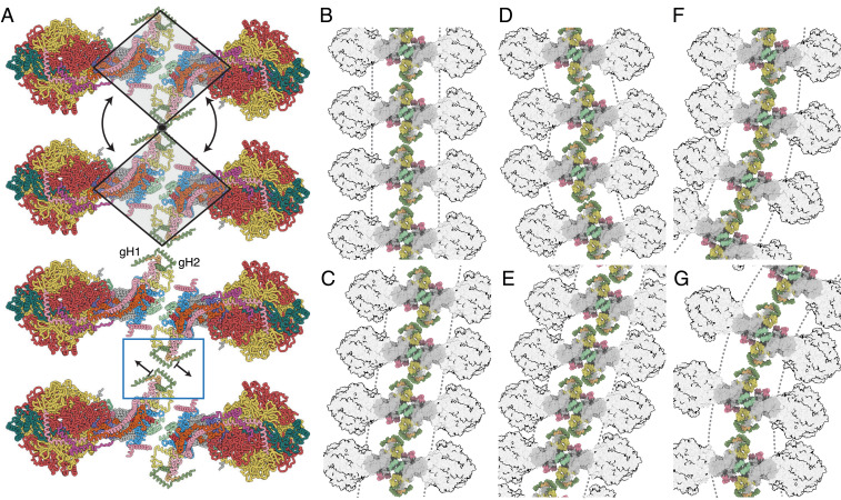
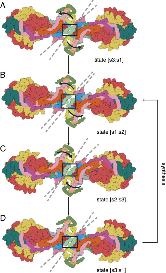
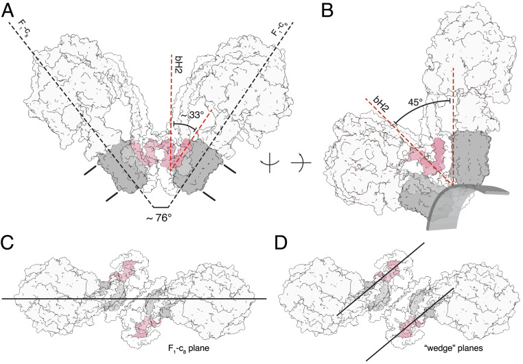
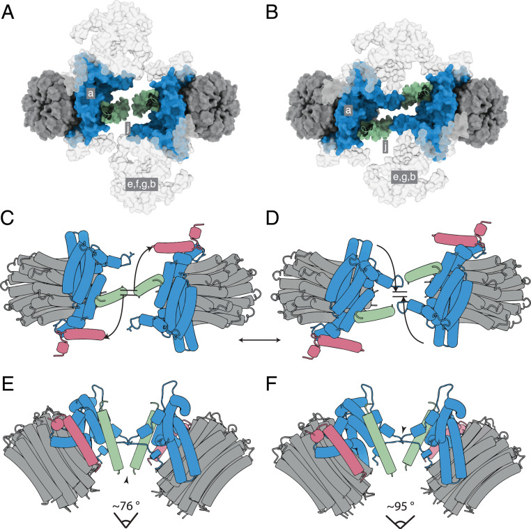
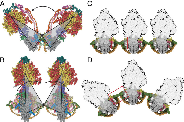
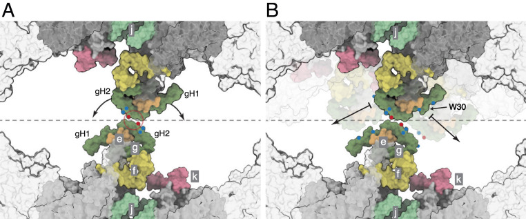
The ATP synthase complexes in mitochondria make the ATP required to sustain life by a rotary mechanism. Their membrane domains are embedded in the inner membranes of the organelle, and they dimerize via interactions between their membrane domains. The dimers form extensive chains along the tips of the cristae with the two rows of monomeric catalytic domains extending into the mitochondrial matrix at an angle to each other. Disruption of the interface between dimers by mutation affects the morphology of the cristae severely. By analysis of particles of purified dimeric bovine ATP synthase by cryo-electron microscopy, we have shown that the angle between the central rotatory axes of the monomeric complexes varies between ca. 76 and 95°. These particles represent active dimeric ATP synthase. Some angular variations arise directly from the catalytic mechanism of the enzyme, and others are independent of catalysis. The monomer-monomer interaction is mediated mainly by j subunits attached to the surface of wedge-shaped protein-lipid structures in the membrane domain of the complex, and the angular variation arises from rotational and translational changes in this interaction, and combinations of both. The structures also suggest how the dimeric ATP synthases might be interacting with each other to form the characteristic rows along the tips of the cristae via other interwedge contacts, molding themselves to the range of oligomeric arrangements observed by tomography of mitochondrial membranes, and at the same time allowing the ATP synthase to operate under the range of physiological conditions that influence the structure of the cristae.

摘要

线粒体中的 ATP 合酶复合物通过旋转机制产生维持生命所需的 ATP。它们的膜结构域嵌入细胞器的内膜中,并通过膜结构域之间的相互作用二聚化。这些二聚体通过在嵴的尖端形成广泛的链,与彼此成一定角度延伸到线粒体基质中的两个单体催化结构域的两排结合在一起。突变破坏二聚体之间的界面会严重影响嵴的形态。通过对纯化的牛二聚体 ATP 合酶颗粒进行冷冻电镜分析,我们表明单体复合物的中心旋转轴之间的角度在约 76 到 95°之间变化。这些颗粒代表活性二聚体 ATP 合酶。一些角度变化直接来自于酶的催化机制,而另一些则与催化无关。单体-单体相互作用主要通过附着在复合物膜结构域中楔形蛋白-脂质结构表面的 j 亚基介导,角度变化来自于这种相互作用的旋转和平移变化,以及两者的组合。这些结构还表明,二聚体 ATP 合酶如何通过其他楔形间接触相互作用,形成沿着嵴尖端的特征行,通过改变自身形状以适应通过线粒体膜断层扫描观察到的寡聚排列范围,同时允许 ATP 合酶在影响嵴结构的一系列生理条件下运行。

https://cdn.ncbi.nlm.nih.gov/pmc/blobs/2024/7923604/5fee83c5da0f/pnas.2021012118fig07.jpg
https://cdn.ncbi.nlm.nih.gov/pmc/blobs/2024/7923604/e22a02789a68/pnas.2021012118fig01.jpg
https://cdn.ncbi.nlm.nih.gov/pmc/blobs/2024/7923604/2c748b6f304a/pnas.2021012118fig02.jpg
https://cdn.ncbi.nlm.nih.gov/pmc/blobs/2024/7923604/41c68803515c/pnas.2021012118fig03.jpg
https://cdn.ncbi.nlm.nih.gov/pmc/blobs/2024/7923604/4716676958c5/pnas.2021012118fig04.jpg
https://cdn.ncbi.nlm.nih.gov/pmc/blobs/2024/7923604/058a9b608d57/pnas.2021012118fig05.jpg
https://cdn.ncbi.nlm.nih.gov/pmc/blobs/2024/7923604/a8f694b189a6/pnas.2021012118fig06.jpg
https://cdn.ncbi.nlm.nih.gov/pmc/blobs/2024/7923604/5fee83c5da0f/pnas.2021012118fig07.jpg

相似文献

1
Interface mobility between monomers in dimeric bovine ATP synthase participates in the ultrastructure of inner mitochondrial membranes.单体间的界面迁移在二聚体牛心 ATP 合酶中参与了线粒体内膜的超微结构。
Proc Natl Acad Sci U S A. 2021 Feb 23;118(8). doi: 10.1073/pnas.2021012118.
2
Structure of the dimeric ATP synthase from bovine mitochondria.牛线粒体二聚体 ATP 合酶的结构。
Proc Natl Acad Sci U S A. 2020 Sep 22;117(38):23519-23526. doi: 10.1073/pnas.2013998117. Epub 2020 Sep 8.
3
Helical arrays of U-shaped ATP synthase dimers form tubular cristae in ciliate mitochondria.U型ATP合酶二聚体的螺旋阵列在纤毛虫线粒体中形成管状嵴。
Proc Natl Acad Sci U S A. 2016 Jul 26;113(30):8442-7. doi: 10.1073/pnas.1525430113. Epub 2016 Jul 11.
4
Structure of a Complete ATP Synthase Dimer Reveals the Molecular Basis of Inner Mitochondrial Membrane Morphology.完整ATP合酶二聚体的结构揭示了线粒体内膜形态的分子基础。
Mol Cell. 2016 Aug 4;63(3):445-56. doi: 10.1016/j.molcel.2016.05.037. Epub 2016 Jun 30.
5
Structure of dimeric ATP synthase from mitochondria: an angular association of monomers induces the strong curvature of the inner membrane.线粒体中二聚体ATP合酶的结构:单体的角向缔合诱导内膜产生强烈弯曲。
FEBS Lett. 2005 Oct 24;579(25):5769-72. doi: 10.1016/j.febslet.2005.09.065. Epub 2005 Oct 6.
6
Dimers of mitochondrial ATP synthase induce membrane curvature and self-assemble into rows.线粒体 ATP 合酶二聚体诱导膜弯曲,并自组装成排。
Proc Natl Acad Sci U S A. 2019 Mar 5;116(10):4250-4255. doi: 10.1073/pnas.1816556116. Epub 2019 Feb 13.
7
How rotating ATP synthases can modulate membrane structure.旋转 ATP 合酶如何调节膜结构。
Arch Biochem Biophys. 2021 Sep 15;708:108939. doi: 10.1016/j.abb.2021.108939. Epub 2021 May 28.
8
Structure of dimeric mitochondrial ATP synthase: novel F0 bridging features and the structural basis of mitochondrial cristae biogenesis.二聚体线粒体ATP合酶的结构:新型F0桥接特征及线粒体嵴生物发生的结构基础
Proc Natl Acad Sci U S A. 2005 Aug 30;102(35):12356-8. doi: 10.1073/pnas.0503893102. Epub 2005 Aug 16.
9
Macromolecular organization of ATP synthase and complex I in whole mitochondria.整个线粒体中 ATP 合酶和复合物 I 的大分子组织。
Proc Natl Acad Sci U S A. 2011 Aug 23;108(34):14121-6. doi: 10.1073/pnas.1103621108. Epub 2011 Aug 11.
10
Cryo-EM of ATP synthases.ATP 合酶的冷冻电镜技术。
Curr Opin Struct Biol. 2018 Oct;52:71-79. doi: 10.1016/j.sbi.2018.08.005. Epub 2018 Sep 18.

引用本文的文献

1
Dominant negative ATP5F1A variants disrupt oxidative phosphorylation causing neurological disorders.显性负性ATP5F1A变体破坏氧化磷酸化,导致神经疾病。
EMBO Mol Med. 2025 Aug 26. doi: 10.1038/s44321-025-00290-8.
2
Cardiolipin acyl chain composition tailors the conformation of mammalian ATP synthase dimers.心磷脂酰基链组成决定了哺乳动物ATP合酶二聚体的构象。
Commun Chem. 2025 Jul 30;8(1):220. doi: 10.1038/s42004-025-01611-1.
3
Dominant negative ATP5F1A variants disrupt oxidative phosphorylation causing neurological disorders.

本文引用的文献

1
Type III ATP synthase is a symmetry-deviated dimer that induces membrane curvature through tetramerization.III 型 ATP 合酶是一种对称性偏离的二聚体,通过四聚化诱导膜弯曲。
Nat Commun. 2020 Oct 22;11(1):5342. doi: 10.1038/s41467-020-18993-6.
2
Cryo-EM structure of the entire mammalian F-type ATP synthase.完整哺乳动物F型ATP合酶的冷冻电镜结构
Nat Struct Mol Biol. 2020 Nov;27(11):1077-1085. doi: 10.1038/s41594-020-0503-8. Epub 2020 Sep 14.
3
Structure of the dimeric ATP synthase from bovine mitochondria.牛线粒体二聚体 ATP 合酶的结构。
显性负性ATP5F1A变体破坏氧化磷酸化,导致神经疾病。
medRxiv. 2025 Jul 8:2025.07.08.25330848. doi: 10.1101/2025.07.08.25330848.
4
Rare Evolutionary Events Support the Phylogenetic Placement of Orthonectida Within Annelida.罕见的进化事件支持直泳虫纲在环节动物门中的系统发育位置。
Int J Mol Sci. 2025 Jun 21;26(13):5983. doi: 10.3390/ijms26135983.
5
Molecular machineries shaping the mitochondrial inner membrane.塑造线粒体内膜的分子机制。
Nat Rev Mol Cell Biol. 2025 May 14. doi: 10.1038/s41580-025-00854-z.
6
High-resolution in situ structures of mammalian respiratory supercomplexes.哺乳动物呼吸超级复合物的高分辨率原位结构。
Nature. 2024 Jul;631(8019):232-239. doi: 10.1038/s41586-024-07488-9. Epub 2024 May 29.
7
Variants in Human ATP Synthase Mitochondrial Genes: Biochemical Dysfunctions, Associated Diseases, and Therapies.人类 ATP 合成酶线粒体基因变异:生化功能障碍、相关疾病和治疗方法。
Int J Mol Sci. 2024 Feb 13;25(4):2239. doi: 10.3390/ijms25042239.
8
The consequence of ATP synthase dimer angle on mitochondrial morphology studied by cryo-electron tomography.通过冷冻电子断层扫描研究ATP合酶二聚体角度对线粒体形态的影响。
Biochem J. 2024 Jan 2;481(3):161-75. doi: 10.1042/BCJ20230450.
9
ATP synthase interactome analysis identifies a new subunit l as a modulator of permeability transition pore in yeast.三磷酸腺苷合酶相互作用组分析鉴定出一个新的亚基 l 作为酵母渗透性转变孔的调节剂。
Sci Rep. 2023 Mar 7;13(1):3839. doi: 10.1038/s41598-023-30966-5.
10
Mitochondrial Cristae Morphology Reflecting Metabolism, Superoxide Formation, Redox Homeostasis, and Pathology.线粒体嵴形态反映代谢、超氧化物形成、氧化还原稳态及病理学
Antioxid Redox Signal. 2023 Oct;39(10-12):635-683. doi: 10.1089/ars.2022.0173. Epub 2023 Apr 11.
Proc Natl Acad Sci U S A. 2020 Sep 22;117(38):23519-23526. doi: 10.1073/pnas.2013998117. Epub 2020 Sep 8.
4
Cryo-EM structures provide insight into how E. coli FF ATP synthase accommodates symmetry mismatch.低温电子显微镜结构提供了对大肠杆菌 FF ATP 合酶如何适应对称失配的深入了解。
Nat Commun. 2020 May 26;11(1):2615. doi: 10.1038/s41467-020-16387-2.
5
The cell biology of mitochondrial membrane dynamics.线粒体膜动力学的细胞生物学。
Nat Rev Mol Cell Biol. 2020 Apr;21(4):204-224. doi: 10.1038/s41580-020-0210-7. Epub 2020 Feb 18.
6
Structure of a mitochondrial ATP synthase with bound native cardiolipin.结合天然心磷脂的线粒体 ATP 合酶的结构。
Elife. 2019 Nov 18;8:e51179. doi: 10.7554/eLife.51179.
7
Individual cristae within the same mitochondrion display different membrane potentials and are functionally independent.同一线粒体中的个体嵴显示出不同的膜电位,并且在功能上是独立的。
EMBO J. 2019 Nov 15;38(22):e101056. doi: 10.15252/embj.2018101056. Epub 2019 Oct 14.
8
Rotary substates of mitochondrial ATP synthase reveal the basis of flexible F-F coupling.线粒体 ATP 合酶的旋转亚基揭示了灵活的 F-F 偶联的基础。
Science. 2019 Jun 21;364(6446). doi: 10.1126/science.aaw9128. Epub 2019 Jun 20.
9
Cryo-EM structure of the mammalian ATP synthase tetramer bound with inhibitory protein IF1.哺乳动物 ATP 合酶四聚体与抑制蛋白 IF1 结合的冷冻电镜结构。
Science. 2019 Jun 14;364(6445):1068-1075. doi: 10.1126/science.aaw4852.
10
Cryo-EM reveals distinct conformations of ATP synthase on exposure to ATP.冷冻电镜揭示了在接触 ATP 时 ATP 合酶的不同构象。
Elife. 2019 Mar 26;8:e43864. doi: 10.7554/eLife.43864.