Sharma Aikta, Goring Alice, Staines Katherine A, Emery Roger J H, Pitsillides Andrew A, Oreffo Richard O C, Mahajan Sumeet, Clarkin Claire E
School of Biological Sciences, Highfield Campus, University of Southampton, Southampton, SO17 1BJ, United Kingdom of Great Britain and Northern Ireland.
School of Applied Sciences, Sighthill Campus, Edinburgh Napier University, Edinburgh, EH11 4BN, United Kingdom of Great Britain and Northern Ireland.
Matrix Biol Plus. 2019 Nov 20;5:100018. doi: 10.1016/j.mbplus.2019.100018. eCollection 2020 Feb.
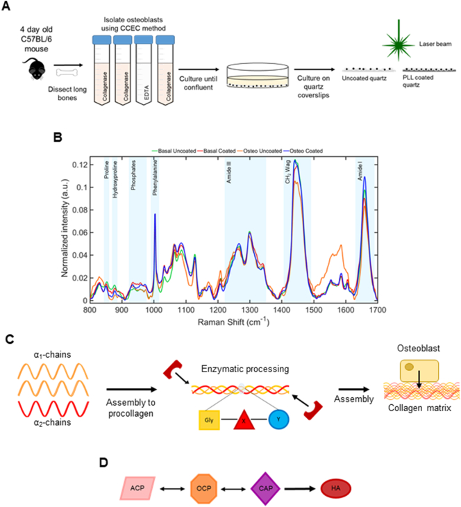
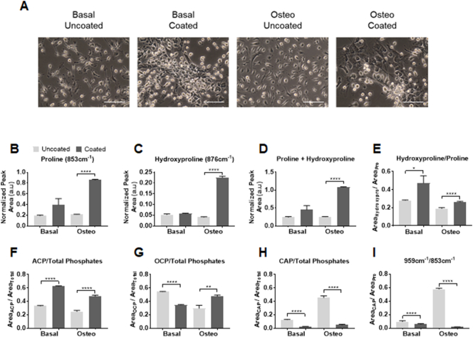
Mineralization of bone is achieved by the sequential maturation of the immature amorphous calcium phase to mature hydroxyapatite (HA) and is central in the process of bone development and repair. To study normal and dysregulated mineralization in vitro, substrates are often coated with poly-l-lysine (PLL) which facilitates cell attachment. This study has used Raman spectroscopy to investigate the effect of PLL coating on osteoblast (OB) matrix composition during differentiation, with a focus on collagen specific proline and hydroxyproline and precursors of HA. Deconvolution analysis of murine derived long bone OB Raman spectra revealed collagen species were 4.01-fold higher in OBs grown on PLL. Further, an increase of 1.91-fold in immature mineral species (amorphous calcium phosphate) was coupled with a 9.32-fold reduction in mature mineral species (carbonated apatite) on PLL versus controls. These unique low mineral signatures identified in OBs were linked with reduced alkaline phosphatase enzymatic activity, reduced Alizarin Red staining and altered osteogenic gene expression. The promotion of immature mineral species and restriction of mature mineral species of OB grown on PLL were linked to increased cell viability and pro-angiogenic vascular endothelial growth factor (VEGF) production. These results demonstrate the utility of Raman spectroscopy to link distinct matrix signatures with OB maturation and VEGF release. Importantly, Raman spectroscopy could provide a label-free approach to clinically assess the angiogenic potential of bone during fracture repair or degenerative bone loss.
骨矿化是通过未成熟的无定形钙相依次成熟为成熟的羟基磷灰石(HA)来实现的,并且在骨发育和修复过程中起着核心作用。为了在体外研究正常和失调的矿化,底物通常用聚-L-赖氨酸(PLL)包被,这有助于细胞附着。本研究利用拉曼光谱研究了PLL包被对成骨细胞(OB)分化过程中基质组成的影响,重点关注胶原蛋白特异性脯氨酸和羟脯氨酸以及HA的前体。对源自小鼠的长骨OB拉曼光谱进行去卷积分析发现,在PLL上生长的OB中胶原蛋白种类高出4.01倍。此外,与对照组相比,PLL上未成熟矿物质种类(无定形磷酸钙)增加了1.91倍,而成熟矿物质种类(碳酸磷灰石)减少了9.32倍。在OB中鉴定出的这些独特的低矿物质特征与碱性磷酸酶活性降低、茜素红染色减少和成骨基因表达改变有关。在PLL上生长的OB中未成熟矿物质种类的增加和成熟矿物质种类的受限与细胞活力增加和促血管生成的血管内皮生长因子(VEGF)产生有关。这些结果证明了拉曼光谱在将不同的基质特征与OB成熟和VEGF释放联系起来方面的实用性。重要的是,拉曼光谱可以提供一种无标记方法,用于临床评估骨折修复或退行性骨质流失期间骨的血管生成潜力。