Section of Surgical Sciences, Vanderbilt University Medical Center, Nashville, Tennessee; Epithelial Biology Center, Vanderbilt University School of Medicine, Nashville, Tennessee.
Department of Cell and Developmental Biology, Vanderbilt University, Nashville, Tennessee.
Cell Mol Gastroenterol Hepatol. 2021;12(1):59-80. doi: 10.1016/j.jcmgh.2021.01.022. Epub 2021 Feb 3.
BACKGROUND & AIMS: The molecular motor, Myosin Vb (MYO5B), is well documented for its role in trafficking cargo to the apical membrane of epithelial cells. Despite its involvement in regulating apical proteins, the role of MYO5B in cell polarity is less clear. Inactivating mutations in MYO5B result in microvillus inclusion disease (MVID), a disorder characterized by loss of key apical transporters and the presence of intracellular inclusions in enterocytes. We previously identified that inclusions in Myo5b knockout (KO) mice form from invagination of the apical brush border via apical bulk endocytosis. Herein, we sought to elucidate the role of polarity complexes and tight junction proteins during the formation of inclusions.
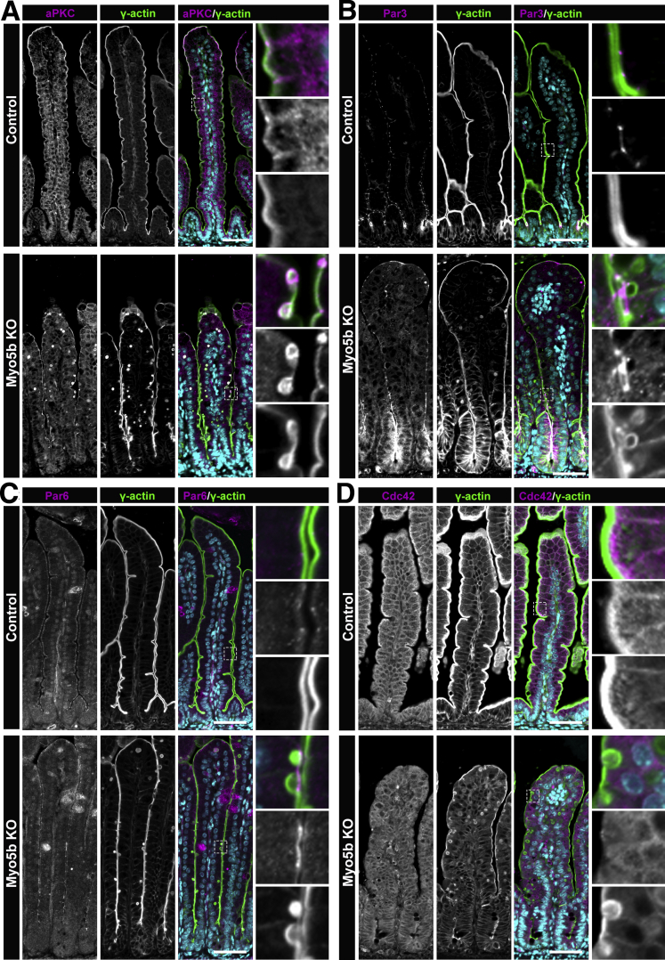
Intestinal tissue from neonatal control and Myo5b KO littermates was analyzed by immunofluorescence to determine the localization of polarity complexes and tight junction proteins.
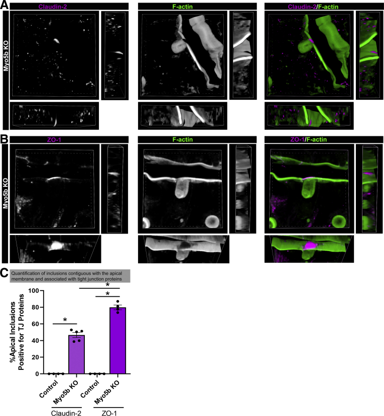
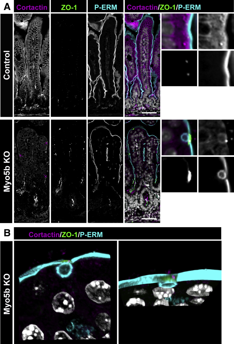
Proteins that make up the apical polarity complexes-Crumbs3 and Pars complexes-were associated with inclusions in Myo5b KO mice. In addition, tight junction proteins were observed to be concentrated over inclusions that were present at the apical membrane of Myo5b-deficient enterocytes in vivo and in vitro. Our mouse findings are complemented by immunostaining in a large animal swine model of MVID genetically engineered to express a human MVID-associated mutation that shows an accumulation of Claudin-2 over forming inclusions. The findings from our swine model of MVID suggest that a similar mechanism of tight junction accumulation occurs in patients with MVID.
These data show that apical bulk endocytosis involves the altered localization of apical polarity proteins and tight junction proteins after loss of Myo5b.
分子马达肌球蛋白 Vb(MYO5B)在将货物运送到上皮细胞顶膜方面的作用已有充分的文献记载。尽管它参与调节顶蛋白,但 MYO5B 在细胞极性中的作用尚不清楚。MYO5B 的失活突变导致微绒毛包涵体病(MVID),这是一种以关键顶质体转运蛋白丢失和肠细胞内包含物存在为特征的疾病。我们之前发现 Myo5b 敲除(KO)小鼠中的包涵体是通过顶膜的顶侧大泡内陷形成的。在此,我们试图阐明在包涵体形成过程中极性复合物和紧密连接蛋白的作用。
通过免疫荧光分析新生对照和 Myo5b KO 同窝仔鼠的肠组织,以确定极性复合物和紧密连接蛋白的定位。
构成顶侧极性复合物的蛋白-Crumbs3 和 Pars 复合物-与 Myo5b KO 小鼠中的包涵体相关。此外,在 Myo5b 缺陷肠细胞的顶膜上存在包涵体时,观察到紧密连接蛋白集中在包涵体上,在体内和体外都是如此。我们在 MVID 的大型动物猪模型中的免疫染色结果补充了这些发现,该模型遗传工程表达了一种与人 MVID 相关突变,该突变显示 Claudin-2 在形成包涵体时的积累。MVID 猪模型的发现表明,在 MVID 患者中,紧密连接的积累可能存在类似的机制。
这些数据表明,在失去 Myo5b 后,顶侧大泡内陷涉及顶侧极性蛋白和紧密连接蛋白的定位改变。