Trembath Dimitri G, Davis Eric S, Rao Shanti, Bradler Evan, Saada Angelica F, Midkiff Bentley R, Snavely Anna C, Ewend Matthew G, Collichio Frances A, Lee Carrie B, Karachaliou Georgia-Sofia, Ayvali Fatih, Ollila David W, Krauze Michal T, Kirkwood John M, Vincent Benjamin G, Nikolaishvilli-Feinberg Nana, Moschos Stergios J
Departments of Pathology and Laboratory Medicine, The University of North Carolina at Chapel Hill, Chapel Hill, NC, United States.
Lineberger Comprehensive Cancer Center, The University of North Carolina at Chapel Hill, Chapel Hill, NC, United States.
Front Oncol. 2021 Jan 21;10:604213. doi: 10.3389/fonc.2020.604213. eCollection 2020.
High tumor-infiltrating lymphocytes (TILs) and hemorrhage are important prognostic factors in patients who have undergone craniotomy for melanoma brain metastases (MBM) before 2011 at the University of Pittsburgh Medical Center (UPMC). We have investigated the prognostic or predictive role of these histopathologic factors in a more contemporary craniotomy cohort from the University of North Carolina at Chapel Hill (UNC-CH). We have also sought to understand better how various immune cell subsets, angiogenic factors, and blood vessels may be associated with clinical and radiographic features in MBM.
Brain tumors from the UPMC and UNC-CH patient cohorts were (re)analyzed by standard histopathology, tumor tissue imaging, and gene expression profiling. Variables were associated with overall survival (OS) and radiographic features.
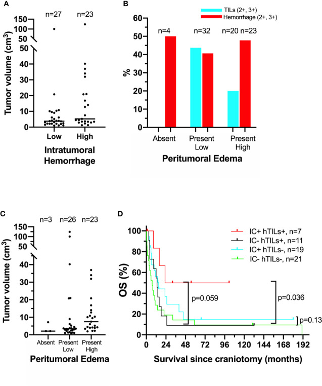
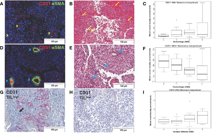
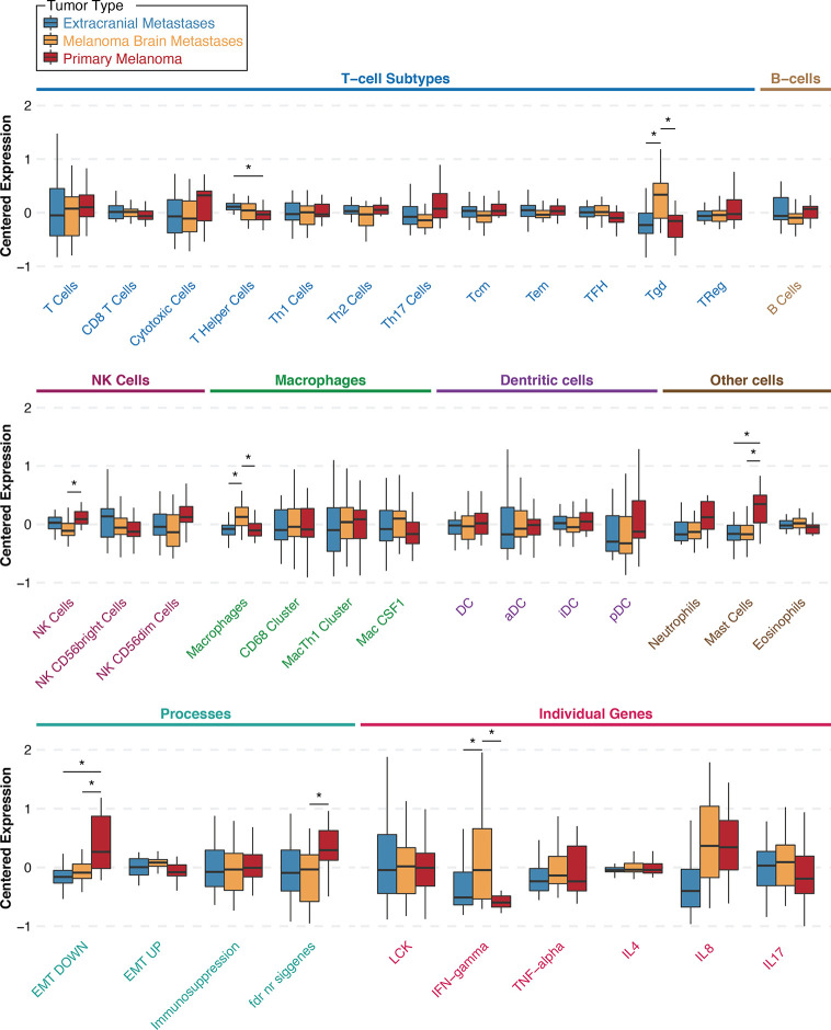
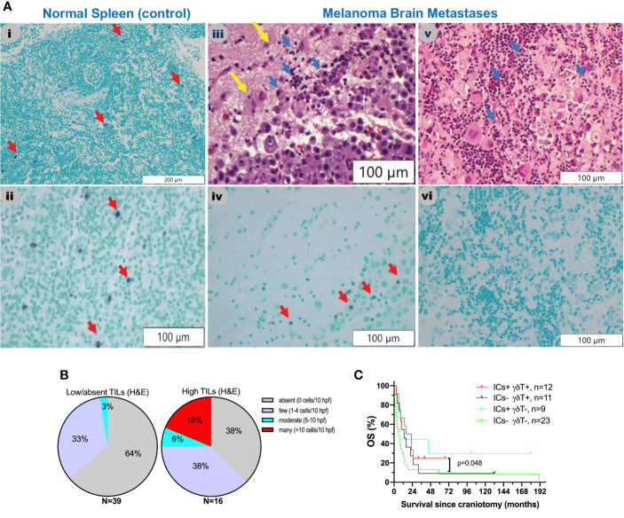
The patient subgroup with high TILs in craniotomy specimens and subsequent treatment with immune checkpoint inhibitors (ICIs, n=7) trended to have longer OS compared to the subgroup with high TILs and no treatment with ICIs (n=11, p=0.059). Bleeding was significantly associated with tumor volume before craniotomy, high melanoma-specific expression of basic fibroblast growth factor (bFGF), and high density of CD31+αSMA- blood vessels. Brain tumors with high versus low peritumoral edema before craniotomy had low (17%) versus high (41%) incidence of brisk TILs. Melanoma-specific expression of the vascular endothelial growth factor (VEGF) was comparable to VEGF expression by TILs and was not associated with any particular prognostic, radiographic, or histopathologic features. A gene signature associated with gamma delta (gd) T cells was significantly higher in intracranial than same-patient extracranial metastases and primary melanoma. However, gdT cell density in MBM was not prognostic.
ICIs may provide greater clinical benefit in patients with brisk TILs in MBM. Intratumoral hemorrhage in brain metastases, a significant clinical problem, is not merely associated with tumor volume but also with underlying biology. bFGF may be an essential pathway to target. VEGF, a factor principally associated with peritumoral edema, is not only produced by melanoma cells but also by TILs. Therefore, suppressing low-grade peritumoral edema using corticosteroids may harm TIL function in 41% of cases. Ongoing clinical trials targeting VEGF in MBM may predict a lack of unfavorable impacts on TIL density and/or intratumoral hemorrhage.
在匹兹堡大学医学中心(UPMC),高肿瘤浸润淋巴细胞(TILs)和出血是2011年前接受开颅手术治疗黑色素瘤脑转移(MBM)患者的重要预后因素。我们在北卡罗来纳大学教堂山分校(UNC-CH)的一个更现代的开颅手术队列中研究了这些组织病理学因素的预后或预测作用。我们还试图更好地了解各种免疫细胞亚群、血管生成因子和血管如何与MBM的临床和影像学特征相关。
对UPMC和UNC-CH患者队列的脑肿瘤进行标准组织病理学、肿瘤组织成像和基因表达谱分析。变量与总生存期(OS)和影像学特征相关。
开颅手术标本中TILs高且随后接受免疫检查点抑制剂(ICI)治疗的患者亚组(n = 7)与TILs高但未接受ICI治疗的亚组(n = 11,p = 0.059)相比,总生存期有延长趋势。出血与开颅手术前的肿瘤体积、碱性成纤维细胞生长因子(bFGF)的黑色素瘤特异性高表达以及CD31 +αSMA-血管的高密度显著相关。开颅手术前瘤周水肿高与低的脑肿瘤中,活跃TILs的发生率分别为41%和17%。血管内皮生长因子(VEGF)的黑色素瘤特异性表达与TILs的VEGF表达相当,且与任何特定的预后、影像学或组织病理学特征均无关联。与γδ(gd)T细胞相关的基因特征在颅内转移瘤中显著高于同一患者的颅外转移瘤和原发性黑色素瘤。然而,MBM中gdT细胞密度无预后意义。
ICI可能在MBM中TILs活跃的患者中提供更大的临床益处。脑转移瘤中的瘤内出血是一个重要的临床问题,不仅与肿瘤体积有关,还与潜在生物学特性有关。bFGF可能是一个关键的靶向途径。VEGF主要与瘤周水肿相关,它不仅由黑色素瘤细胞产生,也由TILs产生。因此,在41%的病例中使用皮质类固醇抑制轻度瘤周水肿可能会损害TIL功能。正在进行的针对MBM中VEGF的临床试验可能预示对TIL密度和/或瘤内出血没有不利影响。