Department of Anesthesiology, University Hospital RWTH Aachen, Pauwelsstrasse 30, 52074 Aachen, Germany.
Institute for Experimental Molecular Imaging, University Hospital RWTH Aachen, Pauwelsstrasse 30, 52074 Aachen, Germany.
Sensors (Basel). 2021 Feb 9;21(4):1200. doi: 10.3390/s21041200.
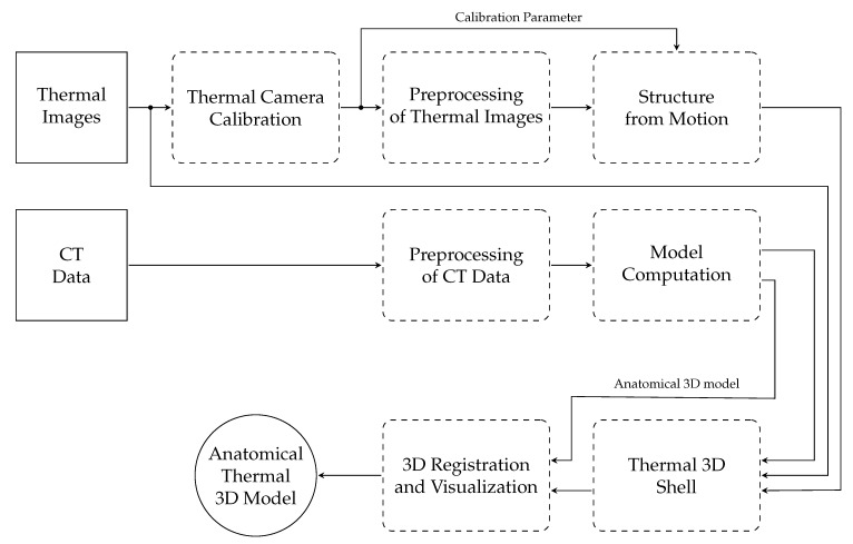
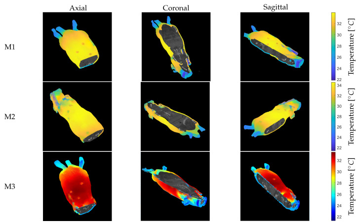
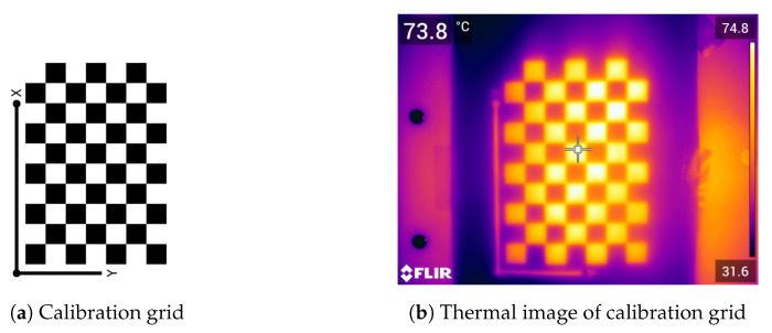
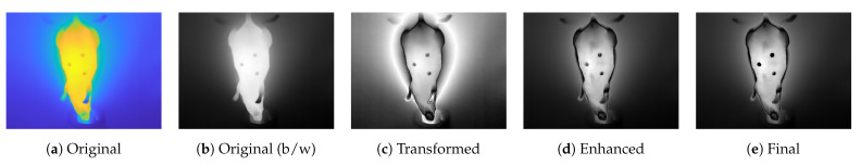
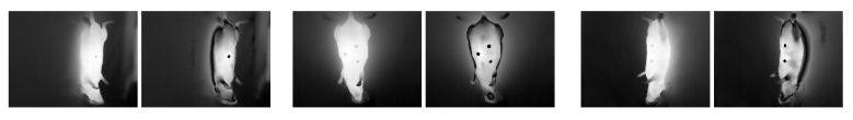
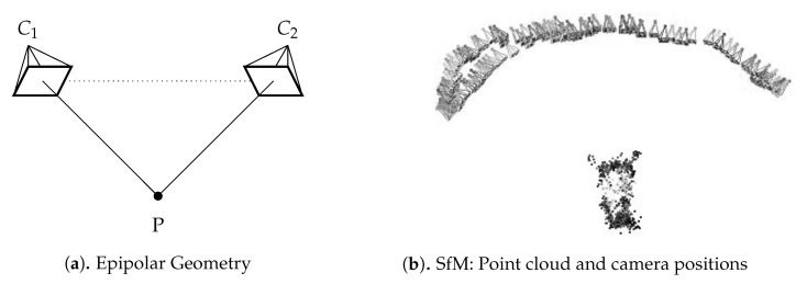
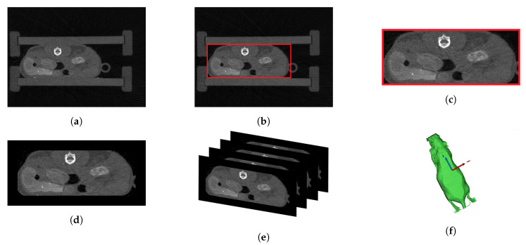
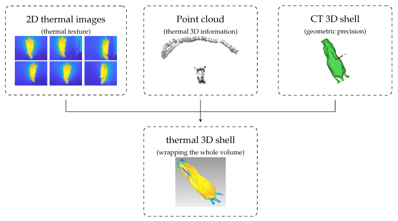
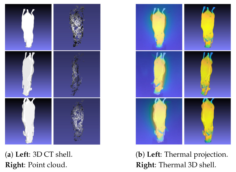
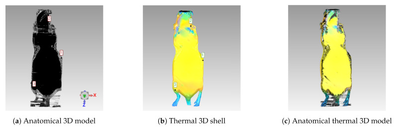
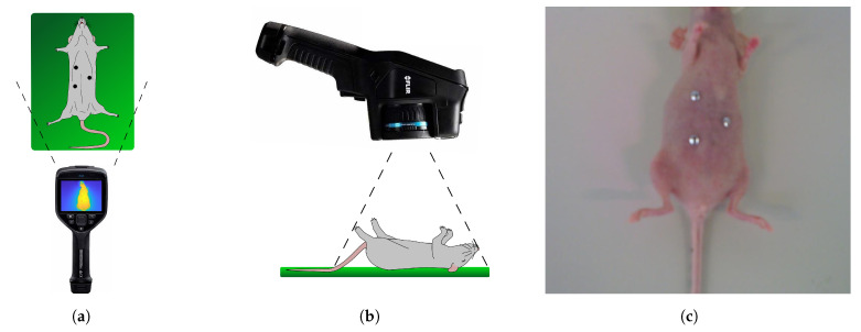
Even though animal trials are a controversial topic, they provide knowledge about diseases and the course of infections in a medical context. To refine the detection of abnormalities that can cause pain and stress to the animal as early as possible, new processes must be developed. Due to its noninvasive nature, thermal imaging is increasingly used for severity assessment in animal-based research. Within a multimodal approach, thermal images combined with anatomical information could be used to simulate the inner temperature profile, thereby allowing the detection of deep-seated infections. This paper presents the generation of anatomical thermal 3D models, forming the underlying multimodal model in this simulation. These models combine anatomical 3D information based on computed tomography (CT) data with a registered thermal shell measured with infrared thermography. The process of generating these models consists of data acquisition (both thermal images and CT), camera calibration, image processing methods, and structure from motion (SfM), among others. Anatomical thermal 3D models were successfully generated using three anesthetized mice. Due to the image processing improvement, the process was also realized for areas with few features, which increases the transferability of the process. The result of this multimodal registration in 3D space can be viewed and analyzed within a visualization tool. Individual CT slices can be analyzed axially, sagittally, and coronally with the corresponding superficial skin temperature distribution. This is an important and successfully implemented milestone on the way to simulating the internal temperature profile. Using this temperature profile, deep-seated infections and inflammation can be detected in order to reduce animal suffering.
尽管动物试验是一个有争议的话题,但它们为医学背景下的疾病和感染过程提供了知识。为了尽早发现可能导致动物疼痛和压力的异常,必须开发新的过程。由于热成像具有非侵入性,因此越来越多地用于动物研究中的严重程度评估。在多模态方法中,可以将热图像与解剖信息结合使用来模拟内部温度分布,从而可以检测深部感染。本文提出了生成解剖学热 3D 模型的方法,该模型构成了该模拟中的基础多模态模型。这些模型将基于计算机断层扫描 (CT) 数据的解剖学 3D 信息与用红外热成像测量的注册热壳结合在一起。生成这些模型的过程包括数据采集(热图像和 CT)、相机校准、图像处理方法和运动结构(SfM)等。成功地使用三只麻醉的老鼠生成了解剖学热 3D 模型。由于图像处理的改进,该过程也可用于特征较少的区域,从而提高了该过程的可转移性。在 3D 空间中的这种多模态配准的结果可以在可视化工具中查看和分析。可以轴向、矢状和冠状地分析各个 CT 切片,并带有相应的表面皮肤温度分布。这是模拟内部温度分布的道路上的一个重要且成功实施的里程碑。使用该温度分布,可以检测深部感染和炎症,以减轻动物的痛苦。