• 文献检索
  • 文档翻译
  • 深度研究
  • 学术资讯
  • Suppr Zotero 插件Zotero 插件
  • 邀请有礼
  • 套餐&价格
  • 历史记录
应用&插件
Suppr Zotero 插件Zotero 插件浏览器插件Mac 客户端Windows 客户端微信小程序
定价
高级版会员购买积分包购买API积分包
服务
文献检索文档翻译深度研究API 文档MCP 服务
关于我们
关于 Suppr公司介绍联系我们用户协议隐私条款
关注我们

Suppr 超能文献

核心技术专利:CN118964589B侵权必究
粤ICP备2023148730 号-1Suppr @ 2026

文献检索

告别复杂PubMed语法,用中文像聊天一样搜索,搜遍4000万医学文献。AI智能推荐,让科研检索更轻松。

立即免费搜索

文件翻译

保留排版,准确专业,支持PDF/Word/PPT等文件格式,支持 12+语言互译。

免费翻译文档

深度研究

AI帮你快速写综述,25分钟生成高质量综述,智能提取关键信息,辅助科研写作。

立即免费体验

HL-1 心脏细胞衍生细胞外囊泡的 microRNA 谱分析。

MicroRNA Profiling of HL-1 Cardiac Cells-Derived Extracellular Vesicles.

机构信息

IRCCS Centro Neurolesi "Bonino-Pulejo", 98124 Messina, Italy.

Department of Innovative Technologies in Medicine & Dentistry, University "G. d'Annunzio", Chieti-Pescara, Via dei Vestini, 31, 66100 Chieti, Italy.

出版信息

Cells. 2021 Jan 30;10(2):273. doi: 10.3390/cells10020273.

DOI:10.3390/cells10020273
PMID:33573156
原文链接:https://pmc.ncbi.nlm.nih.gov/articles/PMC7912193/
Abstract

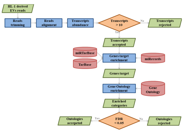
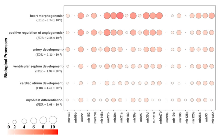
HL-1 is a cell line that shows a phenotype similar to adult cardiomyocytes. All major cardiac cell types release extracellular vesicles (EVs) that emerge as key mediators of intercellular communication. EVs can mediate intercellular cross-talk through the transfer of specific microRNAs (miRNAs). MiRNAs are known to play important regulatory roles during tissue differentiation and regeneration processes. Furthermore, miRNAs have recently been shown to be involved in the proliferation of adult cardiomyocytes. In this context, the purpose of this study was to analyze the transcriptomic profile of miRNAs expressed from HL-1 cardiac muscle cell-derived EVs, using next generation sequencing (NGS). Specifically, our transcriptomic analysis showed that the EVs derived from our HL-1 cells contained miRNAs that induce blood vessel formation and increase cell proliferation. Indeed, our bioinformatics analysis revealed 26 miRNAs expressed in EVs derived from our HL-1 that target genes related to cardiovascular development. In particular, their targets are enriched for the following biological processes related to cardiovascular development: heart morphogenesis, positive regulation of angiogenesis, artery development, ventricular septum development, cardiac atrium development, and myoblast differentiation. Consequently, EVs could become important in the field of regenerative medicine.

摘要

HL-1 细胞系表现出类似于成人心肌细胞的表型。所有主要的心肌细胞类型都会释放细胞外囊泡 (EVs),这些 EVs 成为细胞间通讯的关键介质。EVs 可以通过特定 microRNAs (miRNAs) 的转移来介导细胞间的交流。miRNAs 在组织分化和再生过程中发挥着重要的调节作用。此外,最近的研究表明,miRNAs 参与了成年心肌细胞的增殖。在这种情况下,本研究的目的是使用下一代测序 (NGS) 分析 HL-1 心肌细胞衍生 EVs 中表达的 miRNAs 的转录组谱。具体来说,我们的转录组分析表明,我们从 HL-1 细胞中分离的 EVs 中含有诱导血管形成和增加细胞增殖的 miRNAs。事实上,我们的生物信息学分析显示,我们从 HL-1 细胞中分离的 EVs 中表达了 26 种 miRNA,这些 miRNA 的靶基因与心血管发育有关。特别是,它们的靶基因富集了与心血管发育相关的以下生物学过程:心脏形态发生、血管生成的正调节、动脉发育、心室间隔发育、心房发育和肌母细胞分化。因此,EVs 在再生医学领域可能变得非常重要。

https://cdn.ncbi.nlm.nih.gov/pmc/blobs/4920/7912193/21e28cce78da/cells-10-00273-g003.jpg
https://cdn.ncbi.nlm.nih.gov/pmc/blobs/4920/7912193/cae8377107af/cells-10-00273-g001.jpg
https://cdn.ncbi.nlm.nih.gov/pmc/blobs/4920/7912193/1ac4a0299294/cells-10-00273-g002.jpg
https://cdn.ncbi.nlm.nih.gov/pmc/blobs/4920/7912193/21e28cce78da/cells-10-00273-g003.jpg
https://cdn.ncbi.nlm.nih.gov/pmc/blobs/4920/7912193/cae8377107af/cells-10-00273-g001.jpg
https://cdn.ncbi.nlm.nih.gov/pmc/blobs/4920/7912193/1ac4a0299294/cells-10-00273-g002.jpg
https://cdn.ncbi.nlm.nih.gov/pmc/blobs/4920/7912193/21e28cce78da/cells-10-00273-g003.jpg

相似文献

1
MicroRNA Profiling of HL-1 Cardiac Cells-Derived Extracellular Vesicles.HL-1 心脏细胞衍生细胞外囊泡的 microRNA 谱分析。
Cells. 2021 Jan 30;10(2):273. doi: 10.3390/cells10020273.
2
Extracellular vesicles and microRNAs in the regulation of cardiomyocyte differentiation and proliferation.细胞外囊泡和 microRNAs 在心肌细胞分化和增殖中的调控作用。
Arch Biochem Biophys. 2023 Nov;749:109791. doi: 10.1016/j.abb.2023.109791. Epub 2023 Oct 18.
3
Extracellular Vesicles Derived from Human Gingival Mesenchymal Stem Cells: A Transcriptomic Analysis.人牙龈间充质干细胞来源的细胞外囊泡:转录组学分析。
Genes (Basel). 2020 Jan 21;11(2):118. doi: 10.3390/genes11020118.
4
Cardioprotective role of extracellular vesicles: A highlight on exosome beneficial effects in cardiovascular diseases.细胞外囊泡的心脏保护作用:外泌体对心血管疾病有益作用的重点介绍。
J Cell Physiol. 2019 Dec;234(12):21732-21745. doi: 10.1002/jcp.28894. Epub 2019 May 29.
5
Small RNA deep sequencing discriminates subsets of extracellular vesicles released by melanoma cells--Evidence of unique microRNA cargos.小RNA深度测序可区分黑色素瘤细胞释放的细胞外囊泡亚群——独特微小RNA载荷的证据
RNA Biol. 2015;12(8):810-23. doi: 10.1080/15476286.2015.1056975.
6
Deep Sequencing MicroRNAs from Extracellular Membrane Vesicles Revealed the Association of the Vesicle Cargo with Cellular Origin.从细胞外膜囊泡中深度测序 microRNAs 揭示了囊泡 cargo 与细胞起源的关联。
Int J Mol Sci. 2020 Feb 8;21(3):1141. doi: 10.3390/ijms21031141.
7
Pleiotropic effects of extracellular vesicles from induced pluripotent stem cell-derived cardiomyocytes on ischemic cardiomyopathy: A preclinical study.诱导多能干细胞衍生的心肌细胞外囊泡对缺血性心肌病的多效作用:一项临床前研究。
J Heart Lung Transplant. 2024 Jan;43(1):85-99. doi: 10.1016/j.healun.2023.08.011. Epub 2023 Aug 21.
8
Extracellular Vesicles Secreted by Atherogenic Macrophages Transfer MicroRNA to Inhibit Cell Migration.动脉粥样硬化形成的巨噬细胞分泌的细胞外囊泡将 microRNA 转移到抑制细胞迁移。
Arterioscler Thromb Vasc Biol. 2018 Jan;38(1):49-63. doi: 10.1161/ATVBAHA.117.309795. Epub 2017 Sep 7.
9
Cardiac Extracellular Vesicles in Normal and Infarcted Heart.正常和梗死心脏中的心脏细胞外囊泡
Int J Mol Sci. 2016 Jan 5;17(1):63. doi: 10.3390/ijms17010063.
10
Extracellular vesicular MicroRNA-27a* contributes to cardiac hypertrophy in chronic heart failure.细胞外囊泡 MicroRNA-27a* 促进慢性心力衰竭中的心肌肥厚。
J Mol Cell Cardiol. 2020 Jun;143:120-131. doi: 10.1016/j.yjmcc.2020.04.032. Epub 2020 May 1.

引用本文的文献

1
Exosome-mediated effects of BRCA1 on cardiovascular artery disease.BRCA1通过外泌体介导对心血管疾病的影响。
Cell Biol Toxicol. 2025 Mar 13;41(1):59. doi: 10.1007/s10565-025-09996-4.
2
Decreased gut microbiome-derived indole-3-propionic acid mediates the exacerbation of myocardial ischemia/reperfusion injury following depression via the brain-gut-heart axis.肠道微生物群衍生的吲哚-3-丙酸减少通过脑-肠-心轴介导抑郁症后心肌缺血/再灌注损伤的加重。
Redox Biol. 2025 Apr;81:103580. doi: 10.1016/j.redox.2025.103580. Epub 2025 Mar 5.
3
Thyroid Hormone Upregulates Cav1.2 Channels in Cardiac Cells via the Downregulation of the Channels' β4 Subunit.

本文引用的文献

1
Mir-30d Regulates Cardiac Remodeling by Intracellular and Paracrine Signaling.miR-30d 通过细胞内和旁分泌信号调节心脏重塑。
Circ Res. 2021 Jan 8;128(1):e1-e23. doi: 10.1161/CIRCRESAHA.120.317244. Epub 2020 Oct 22.
2
MicroRNA-183 as a Novel Regulator Protects Against Cardiomyocytes Hypertrophy via Targeting TIAM1.微小 RNA-183 作为一种新型调节因子,通过靶向 TIAM1 保护心肌细胞肥大。
Am J Hypertens. 2022 Jan 5;35(1):87-95. doi: 10.1093/ajh/hpaa144.
3
miR-378a-3p inhibits ischemia/reperfusion-induced apoptosis in H9C2 cardiomyocytes by targeting TRIM55 via the DUSP1-JNK1/2 signaling pathway.
甲状腺激素通过下调通道β4 亚基上调心肌细胞中的 Cav1.2 通道。
Int J Mol Sci. 2024 Oct 8;25(19):10798. doi: 10.3390/ijms251910798.
4
Exosomes in Atherosclerosis: Role in the Pathogenesis and Targets for Therapy.动脉粥样硬化中的外泌体:在发病机制中的作用及治疗靶点
Curr Med Chem. 2024 May 3. doi: 10.2174/0109298673302220240430173404.
5
Endothelial Extracellular Vesicles Enriched in microRNA-34a Predict New-Onset Diabetes in Coronavirus Disease 2019 (COVID-19) Patients: Novel Insights for Long COVID Metabolic Sequelae.富含微小RNA-34a的内皮细胞外囊泡可预测2019冠状病毒病(COVID-19)患者的新发糖尿病:对新冠后代谢后遗症的新见解
J Pharmacol Exp Ther. 2024 Mar 15;389(1):34-39. doi: 10.1124/jpet.122.001253.
6
Immune cell-derived extracellular vesicular microRNAs induce pancreatic beta cell apoptosis.免疫细胞衍生的细胞外囊泡微小RNA诱导胰腺β细胞凋亡。
Heliyon. 2022 Dec 2;8(12):e11995. doi: 10.1016/j.heliyon.2022.e11995. eCollection 2022 Dec.
miR-378a-3p 通过靶向 TRIM55 抑制 DUSP1-JNK1/2 信号通路抑制 H9C2 心肌细胞缺血/再灌注诱导的细胞凋亡。
Aging (Albany NY). 2020 May 28;12(10):8939-8952. doi: 10.18632/aging.103106.
4
MicroRNA-27 attenuates pressure overload-Induced cardiac hypertrophy and dysfunction by targeting galectin-3.MicroRNA-27 通过靶向半乳糖凝集素-3 减轻压力超负荷诱导的心肌肥厚和功能障碍。
Arch Biochem Biophys. 2020 Aug 15;689:108405. doi: 10.1016/j.abb.2020.108405. Epub 2020 May 18.
5
miR-25 Promotes Cardiomyocyte Proliferation by Targeting FBXW7.微小RNA-25通过靶向F-box蛋白7促进心肌细胞增殖。
Mol Ther Nucleic Acids. 2020 Mar 6;19:1299-1308. doi: 10.1016/j.omtn.2020.01.013. Epub 2020 Jan 21.
6
Characterization of human cardiac mesenchymal stromal cells and their extracellular vesicles comparing with human bone marrow derived mesenchymal stem cells.比较人心肌间充质基质细胞及其细胞外囊泡与人骨髓间充质干细胞的特征。
BMB Rep. 2020 Feb;53(2):118-123. doi: 10.5483/BMBRep.2020.53.2.235.
7
MircroRNA-10b Promotes Human Embryonic Stem Cell-Derived Cardiomyocyte Proliferation via Novel Target Gene LATS1.微小RNA-10b通过新靶基因LATS1促进人胚胎干细胞衍生的心肌细胞增殖。
Mol Ther Nucleic Acids. 2020 Mar 6;19:437-445. doi: 10.1016/j.omtn.2019.11.026. Epub 2019 Nov 29.
8
microRNA-451a prevents activation of matrix metalloproteinases 2 and 9 in human cardiomyocytes during pathological stress stimulation.microRNA-451a 可防止人心肌细胞在病理应激刺激期间激活基质金属蛋白酶 2 和 9。
Am J Physiol Cell Physiol. 2020 Jan 1;318(1):C94-C102. doi: 10.1152/ajpcell.00204.2019. Epub 2019 Oct 16.
9
MiRNA-182 regulates the cardiomyocyte apoptosis in heart failure.miRNA-182 调控心力衰竭中心肌细胞凋亡。
Eur Rev Med Pharmacol Sci. 2019 Jun;23(11):4917-4923. doi: 10.26355/eurrev_201906_18079.
10
Advances in Technologies for Purification and Enrichment of Extracellular Vesicles.用于细胞外囊泡的纯化和富集的技术进展。
SLAS Technol. 2019 Oct;24(5):477-488. doi: 10.1177/2472630319846877. Epub 2019 May 14.