Department of Chemistry and Biochemistry, Mendel University in Brno, Zemedelska 1, CZ-613 00 Brno, Czech Republic.
Laboratory of Bioimaging and Pathologies, CNRS UMR 7021, Faculty of Pharmacy, Strasbourg University, 74, route du Rhin, F-67401 Illkirch, France.
Viruses. 2021 Jan 30;13(2):213. doi: 10.3390/v13020213.
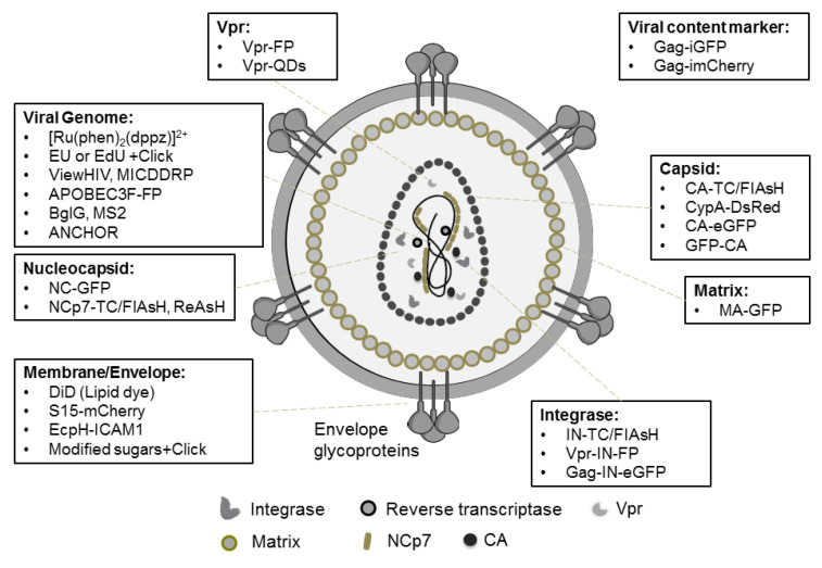
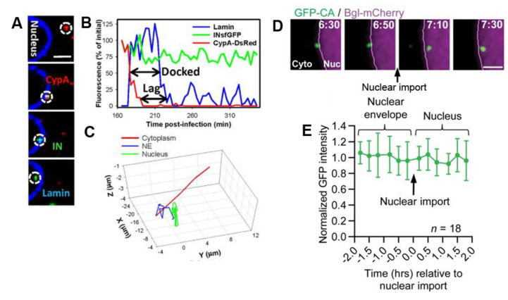
During the last two decades, progresses in bioimaging and the development of various strategies to fluorescently label the viral components opened a wide range of possibilities to visualize the early phase of Human Immunodeficiency Virus 1 (HIV-1) life cycle directly in infected cells. After fusion of the viral envelope with the cell membrane, the viral core is released into the cytoplasm and the viral RNA (vRNA) is retro-transcribed into DNA by the reverse transcriptase. During this process, the RNA-based viral complex transforms into a pre-integration complex (PIC), composed of the viral genomic DNA (vDNA) coated with viral and host cellular proteins. The protective capsid shell disassembles during a process called uncoating. The viral genome is transported into the cell nucleus and integrates into the host cell chromatin. Unlike biochemical approaches that provide global data about the whole population of viral particles, imaging techniques enable following individual viruses on a single particle level. In this context, quantitative microscopy has brought original data shedding light on the dynamics of the viral entry into the host cell, the cytoplasmic transport, the nuclear import, and the selection of the integration site. In parallel, multi-color imaging studies have elucidated the mechanism of action of host cell factors implicated in HIV-1 viral cycle progression. In this review, we describe the labeling strategies used for HIV-1 fluorescence imaging and report on the main advancements that imaging studies have brought in the understanding of the infection mechanisms from the viral entry into the host cell until the provirus integration step.
在过去的二十年中,生物成像技术的进步和各种荧光标记病毒成分的策略的发展,为直接在感染细胞中可视化人类免疫缺陷病毒 1(HIV-1)生命周期的早期阶段提供了广泛的可能性。病毒包膜与细胞膜融合后,病毒核心被释放到细胞质中,逆转录酶将病毒 RNA(vRNA)逆转录成 DNA。在此过程中,基于 RNA 的病毒复合物转化为前整合复合物(PIC),由包裹病毒和宿主细胞蛋白的病毒基因组 DNA(vDNA)组成。在称为脱壳的过程中,保护性衣壳壳解体。病毒基因组被运送到细胞核并整合到宿主细胞染色质中。与提供有关整个病毒颗粒群体的全局数据的生化方法不同,成像技术能够在单个颗粒水平上跟踪单个病毒。在这种情况下,定量显微镜带来了原始数据,揭示了病毒进入宿主细胞、细胞质运输、核输入以及整合位点选择的动力学。同时,多色成像研究阐明了宿主细胞因子在 HIV-1 病毒周期进展中作用的机制。在这篇综述中,我们描述了用于 HIV-1 荧光成像的标记策略,并报告了成像研究在理解从病毒进入宿主细胞到前病毒整合步骤的感染机制方面带来的主要进展。