Wirth Stephan H, Rahm Stefan, Kamath Atul F, Dora Claudio, Zingg Patrick O
Department of Orthopaedics, University of Zürich, Balgrist Hospital, Zürich 8032, Switzerland.
Center for Hip Preservation, Orthopaedic and Rheumatologic Institute, Cleveland Clinic, Cleveland, OH 44139, USA.
J Hip Preserv Surg. 2019 Oct 24;6(4):411-420. doi: 10.1093/jhps/hnz051. eCollection 2019 Dec.
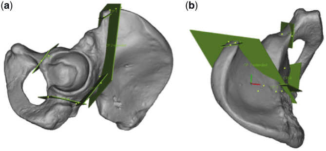
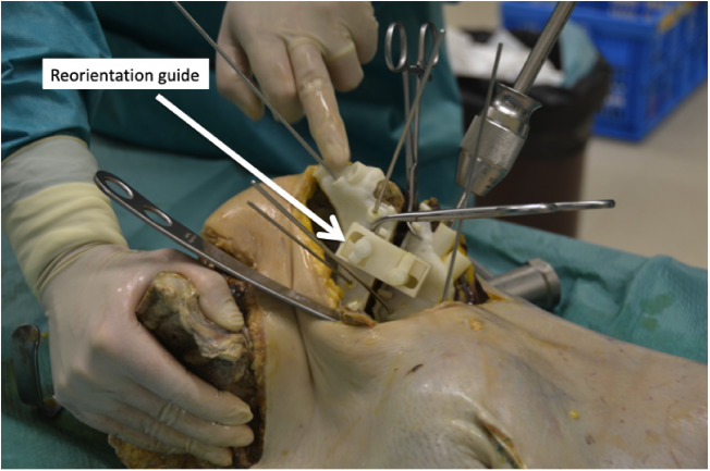
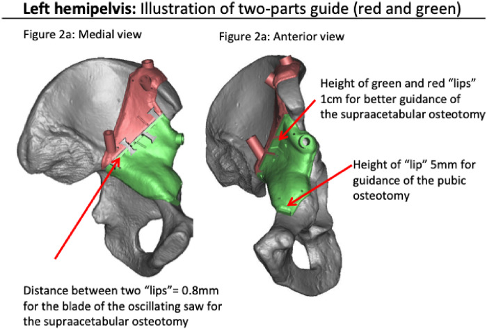
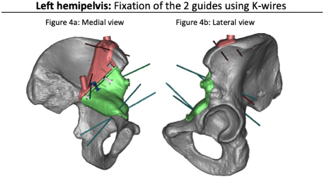
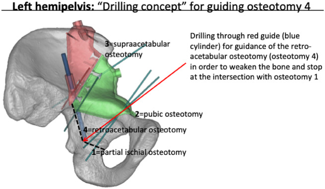
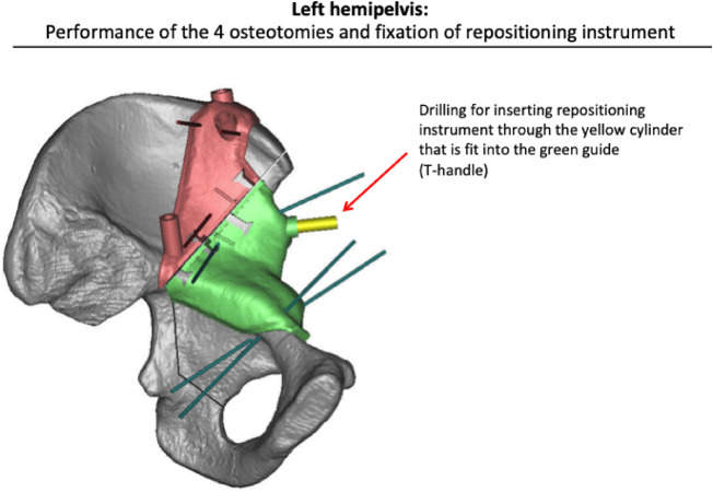
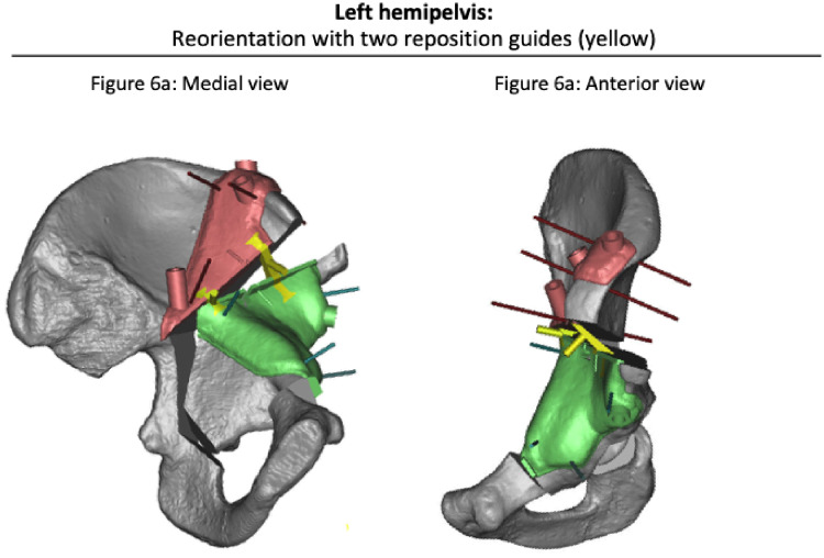
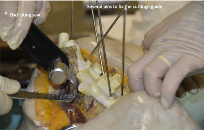
The goal of periacetabular osteotomy (PAO) is to reorient the acetabulum in a more physiological position. Its realization remains challenging regarding the final position of the acetabulum. Assistance with custom cutting- and reorientation-guides would thus be very helpful. Our purpose is to present a pilot study on such guides. Eight cadaveric hemipelvis were scanned using CT. After segmentation, 3D models of each specimen were created, a PAO was virtually performed and reorientation of the acetabula were defined. A specific guide was designed aiming to assist in iliac, posterior column and superior pubic ramus cuts as well as in acetabulum reorientation. Furthermore, the acetabular position was planned. Three-dimensional printed guides were used to perform PAO using the modified Smith-Peterson approach. The post-operative CT images and virtually planned acetabulum reorientation were compared in terms of acetabular index (AC), lateral centre edge angle (LCE), acetabular anteversion angle (AcetAV). There was no intra-articular or posterior column fracture seen. Two cadavers showed very low bone quality with insufficient stability of fixation and were excluded from further analysis. Correlation between the post-operative result and planning of the six included cadavers revealed the following mean deviations: 5° (SD ±3°) for AC angle, 6° (SD ±4°) for LCE angle and 15° (SD ±11°) for AcetAV angle. The use of 3D cutting and reorientation blocks for PAO was possible through a modified Smith-Peterson approach and revealed accurate fit to bone, accurate positioning of the osteotomies and acceptable planned corrections in cadavers with good bone quality.
髋臼周围截骨术(PAO)的目标是将髋臼重新定位到更符合生理的位置。在髋臼的最终位置方面,实现这一目标仍然具有挑战性。因此,使用定制的切割和重新定位导向器将非常有帮助。我们的目的是展示一项关于此类导向器的初步研究。使用CT扫描了8个尸体半骨盆。分割后,创建了每个标本的三维模型,虚拟执行了PAO并定义了髋臼的重新定位。设计了一种特定的导向器,旨在辅助进行髂骨、后柱和耻骨上支的切割以及髋臼的重新定位。此外,还规划了髋臼的位置。使用改良的Smith-Peterson方法,使用三维打印导向器进行PAO。比较了术后CT图像和虚拟规划的髋臼重新定位在髋臼指数(AC)、外侧中心边缘角(LCE)、髋臼前倾角(AcetAV)方面的情况。未发现关节内或后柱骨折。有两具尸体显示骨质非常低,固定稳定性不足,被排除在进一步分析之外。对纳入的6具尸体的术后结果与规划之间的相关性显示出以下平均偏差:AC角为5°(标准差±3°),LCE角为6°(标准差±4°),AcetAV角为15°(标准差±11°)。通过改良的Smith-Peterson方法,使用三维切割和重新定位块进行PAO是可行的,并且在骨质良好的尸体中显示出与骨骼的精确贴合、截骨的精确定位以及可接受的规划矫正。