Hoch Armando, Roth Tabitha, Marcon Magda, Fürnstahl Philipp, Fucentese Sandro F, Sutter Reto
Department of Orthopaedics, Balgrist University Hospital, University of Zurich, Forchstrasse 340, 8008, Zurich, Switzerland.
Research in Orthopaedic Computer Science, Balgrist University Hospital, University of Zurich, Zurich, Switzerland.
Insights Imaging. 2021 Feb 15;12(1):18. doi: 10.1186/s13244-020-00960-w.
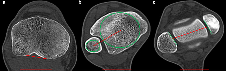
Pathological tibial torsion is known to negatively influence the functionality of the lower extremity, and therefore, its assessment might play an important role. While 3D imaging is used for many examinations of the musculoskeletal system, for the determination of tibial torsion no 3D measurement technique has been available so far. We developed a 3D measurement method and assess its interobserver reliability as well as its correlation with standard 2D measurement methods.
CT scans of 82 tibiae in 79 patients with a mean age of 41 years were included. A novel 3D measurement technique was developed and applied. Measurements were compared with two frequently used 2D measurement methods. ICC (intraclass correlation coefficient) for the new technique was determined and compared to the 2D measurement method. Furthermore, differences between left and right legs as well as between males and females were assessed.
The ICC for the 2D methods was 0.917 and 0.938, respectively. For the 3D measurements, ICCs were calculated to be 0.954 and 0.950. Agreement between 2 and 3D methods was moderate to good with ICCs between 0.715 and 0.795. Torsion values for left and right legs did not differ significantly in 2D and in 3D (26.2 vs 28.5° and 27.2 vs. 25.9°). The same is true for the differences between male and female in 2D and 3D (26.2 vs. 29.6° and 25.0 vs. 31.2°).
The newly developed 3D measurement technique shows a high intraclass agreement and offers an applicable opportunity to assess the tibial torsion three-dimensionally.
病理性胫骨扭转已知会对下肢功能产生负面影响,因此,对其进行评估可能具有重要作用。虽然三维成像被用于肌肉骨骼系统的许多检查,但到目前为止,尚无用于确定胫骨扭转的三维测量技术。我们开发了一种三维测量方法,并评估其观察者间的可靠性以及与标准二维测量方法的相关性。
纳入79例平均年龄41岁患者的82例胫骨的CT扫描图像。开发并应用了一种新的三维测量技术。将测量结果与两种常用的二维测量方法进行比较。确定新技术的组内相关系数(ICC)并与二维测量方法进行比较。此外,评估左右腿之间以及男女之间的差异。
二维方法的ICC分别为0.917和0.938。对于三维测量,计算得出的ICC为0.954和0.950。二维和三维方法之间的一致性为中等至良好,ICC在0.715和0.795之间。二维和三维测量中左右腿的扭转值无显著差异(26.2°对28.5°以及27.2°对25.9°)。二维和三维测量中男女之间的差异情况相同(26.2°对29.6°以及25.0°对31.2°)。
新开发的三维测量技术显示出较高的组内一致性,并为三维评估胫骨扭转提供了一种可行的方法。