Department of Diagnostic and Interventional Radiology, University Hospital Düsseldorf, Düsseldorf, Germany.
Institute of Imaging and Computer Vision, RWTH University Aachen, Aachen, Germany.
Sci Rep. 2021 Dec 1;11(1):23244. doi: 10.1038/s41598-021-02708-y.
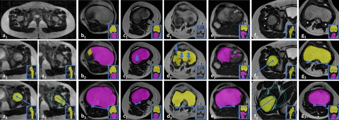
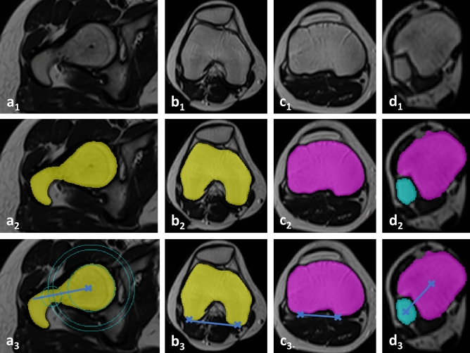
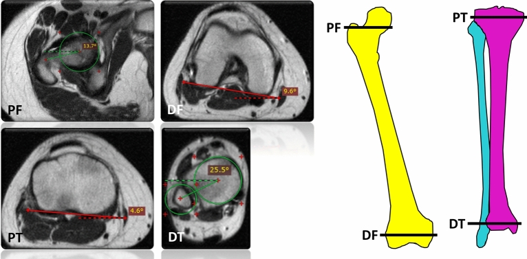
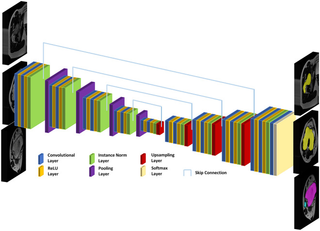
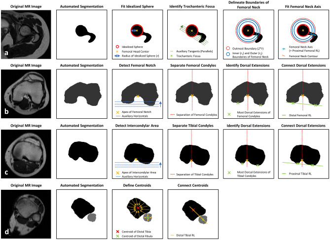
Abnormal torsion of the lower limbs may adversely affect joint health. This study developed and validated a deep learning-based method for automatic measurement of femoral and tibial torsion on MRI. Axial T2-weighted sequences acquired of the hips, knees, and ankles of 93 patients (mean age, 13 ± 5 years; 52 males) were included and allocated to training (n = 60), validation (n = 9), and test sets (n = 24). A U-net convolutional neural network was trained to segment both femur and tibia, identify osseous anatomic landmarks, define pertinent reference lines, and quantify femoral and tibial torsion. Manual measurements by two radiologists provided the reference standard. Inter-reader comparisons were performed using repeated-measures ANOVA, Pearson's r, and the intraclass correlation coefficient (ICC). Mean Sørensen-Dice coefficients for segmentation accuracy ranged between 0.89 and 0.93 and erroneous segmentations were scarce. Ranges of torsion as measured by both readers and the algorithm on the same axial image were 15.8°-18.0° (femur) and 33.9°-35.2° (tibia). Correlation coefficients (ranges, .968 ≤ r ≤ .984 [femur]; .867 ≤ r ≤ .904 [tibia]) and ICCs (ranges, .963 ≤ ICC ≤ .974 [femur]; .867 ≤ ICC ≤ .894 [tibia]) indicated excellent inter-reader agreement. Algorithm-based analysis was faster than manual analysis (7 vs 207 vs 230 s, p < .001). In conclusion, fully automatic measurement of torsional alignment is accurate, reliable, and sufficiently fast for clinical workflows.
下肢异常扭转可能会对关节健康产生不利影响。本研究开发并验证了一种基于深度学习的方法,用于自动测量 MRI 上的股骨和胫骨扭转。纳入了 93 名患者(平均年龄 13 ± 5 岁;52 名男性)的髋关节、膝关节和踝关节的轴向 T2 加权序列,并将其分配到训练集(n=60)、验证集(n=9)和测试集(n=24)。使用 U 型网络卷积神经网络对股骨和胫骨进行分割,识别骨性解剖标志,定义相关参考线,并量化股骨和胫骨扭转。两名放射科医生的手动测量值作为参考标准。采用重复测量方差分析、Pearson r 和组内相关系数(ICC)进行读者间比较。分割准确性的平均 Sørensen-Dice 系数在 0.89 到 0.93 之间,错误分割很少。两位读者和算法在同一轴向图像上测量的扭转范围分别为 15.8°-18.0°(股骨)和 33.9°-35.2°(胫骨)。相关系数(范围,0.968≤r≤0.984 [股骨];0.867≤r≤0.904 [胫骨])和 ICC(范围,0.963≤ICC≤0.974 [股骨];0.867≤ICC≤0.894 [胫骨])表明读者间具有极好的一致性。基于算法的分析比手动分析更快(7 秒对 207 秒对 230 秒,p<0.001)。总之,用于测量扭转对线的全自动方法准确、可靠,并且分析速度足够满足临床工作流程的需求。