• 文献检索
  • 文档翻译
  • 深度研究
  • 学术资讯
  • Suppr Zotero 插件Zotero 插件
  • 邀请有礼
  • 套餐&价格
  • 历史记录
应用&插件
Suppr Zotero 插件Zotero 插件浏览器插件Mac 客户端Windows 客户端微信小程序
定价
高级版会员购买积分包购买API积分包
服务
文献检索文档翻译深度研究API 文档MCP 服务
关于我们
关于 Suppr公司介绍联系我们用户协议隐私条款
关注我们

Suppr 超能文献

核心技术专利:CN118964589B侵权必究
粤ICP备2023148730 号-1Suppr @ 2026

文献检索

告别复杂PubMed语法,用中文像聊天一样搜索,搜遍4000万医学文献。AI智能推荐,让科研检索更轻松。

立即免费搜索

文件翻译

保留排版,准确专业,支持PDF/Word/PPT等文件格式,支持 12+语言互译。

免费翻译文档

深度研究

AI帮你快速写综述,25分钟生成高质量综述,智能提取关键信息,辅助科研写作。

立即免费体验

临床实践中的共聚焦显微镜检查。

Specular microscopy in clinical practice.

机构信息

Cornea and Anterior Segment Services, LV Prasad Eye Institute, Hyderabad, Telangana, India.

Cornea, Cataract and Refractive Surgery Service, RP Centre, AIIMS, New Delhi, India.

出版信息

Indian J Ophthalmol. 2021 Mar;69(3):517-524. doi: 10.4103/ijo.IJO_574_20.

DOI:10.4103/ijo.IJO_574_20
PMID:33595465
原文链接:https://pmc.ncbi.nlm.nih.gov/articles/PMC7942069/
Abstract

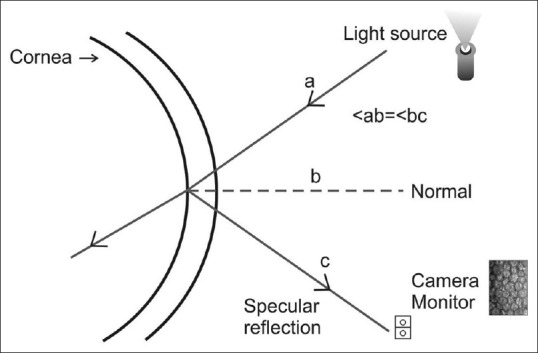
Specular microscopy is a noninvasive diagnostic tool that allows for in vivo evaluation of corneal endothelium in health and various diseased states. Endothelial imaging helps in the diagnosis and management of several endothelial disorders. The review focuses on the principles of specular microscopy, limitations of endothelial imaging, and its interpretation in common conditions seen in the clinical practice. A thorough PubMed search was done using the keywords specular microscopy, corneal endothelium, and endothelial imaging.

摘要

共聚焦显微镜是一种非侵入性的诊断工具,可用于在体内评估健康和各种病变状态下的角膜内皮。内皮成像有助于诊断和治疗几种内皮疾病。本文综述了共焦显微镜的原理、内皮成像的局限性及其在临床常见疾病中的解读。使用关键词共焦显微镜、角膜内皮和内皮成像,对 PubMed 进行了全面检索。

https://cdn.ncbi.nlm.nih.gov/pmc/blobs/ca72/7942069/6478ce62688b/IJO-69-517-g014.jpg
https://cdn.ncbi.nlm.nih.gov/pmc/blobs/ca72/7942069/3b096756f7de/IJO-69-517-g001.jpg
https://cdn.ncbi.nlm.nih.gov/pmc/blobs/ca72/7942069/60b360d3ccd4/IJO-69-517-g002.jpg
https://cdn.ncbi.nlm.nih.gov/pmc/blobs/ca72/7942069/38b2380cd1e3/IJO-69-517-g005.jpg
https://cdn.ncbi.nlm.nih.gov/pmc/blobs/ca72/7942069/a637cfa6eb47/IJO-69-517-g006.jpg
https://cdn.ncbi.nlm.nih.gov/pmc/blobs/ca72/7942069/34fda36ae40f/IJO-69-517-g007.jpg
https://cdn.ncbi.nlm.nih.gov/pmc/blobs/ca72/7942069/fba397902f30/IJO-69-517-g008.jpg
https://cdn.ncbi.nlm.nih.gov/pmc/blobs/ca72/7942069/05351e3d5591/IJO-69-517-g009.jpg
https://cdn.ncbi.nlm.nih.gov/pmc/blobs/ca72/7942069/50ecc3fd4e4c/IJO-69-517-g010.jpg
https://cdn.ncbi.nlm.nih.gov/pmc/blobs/ca72/7942069/0301da070d62/IJO-69-517-g011.jpg
https://cdn.ncbi.nlm.nih.gov/pmc/blobs/ca72/7942069/18b7f59571b4/IJO-69-517-g012.jpg
https://cdn.ncbi.nlm.nih.gov/pmc/blobs/ca72/7942069/95827cacabf4/IJO-69-517-g013.jpg
https://cdn.ncbi.nlm.nih.gov/pmc/blobs/ca72/7942069/6478ce62688b/IJO-69-517-g014.jpg

相似文献

1
Specular microscopy in clinical practice.临床实践中的共聚焦显微镜检查。
Indian J Ophthalmol. 2021 Mar;69(3):517-524. doi: 10.4103/ijo.IJO_574_20.
2
Specular Microscopy镜面显微镜检查
3
Comparison of confocal biomicroscopy and noncontact specular microscopy for evaluation of the corneal endothelium.共焦生物显微镜检查与非接触式角膜内皮显微镜检查在评估角膜内皮方面的比较。
Cornea. 2003 Aug;22(6):512-5. doi: 10.1097/00003226-200308000-00005.
4
Endothelium and pachymetry of clear corneal grafts 15 to 33 years after penetrating keratoplasty.穿透性角膜移植术后15至33年透明角膜移植片的内皮和测厚
Am J Ophthalmol. 1999 May;127(5):600-2. doi: 10.1016/s0002-9394(98)00406-1.
5
Corneal endothelial cell density and pachymetry measured by contact and noncontact specular microscopy.通过接触式和非接触式镜面显微镜测量角膜内皮细胞密度和角膜厚度。
J Cataract Refract Surg. 2002 Oct;28(10):1763-9. doi: 10.1016/s0886-3350(02)01296-8.
6
Significant differences between specular microscopy and corneal bank endothelial cell counts - a pilot study.共焦激光显微镜和角膜银行内皮细胞计数的显著差异-一项初步研究。
Acta Ophthalmol. 2019 Dec;97(8):e1077-e1081. doi: 10.1111/aos.14185. Epub 2019 Jul 8.
7
Specular microscopy of the corneal endothelial cells of bovines: an study.牛眼角膜内皮细胞的共焦显微镜观察:一项研究。
Open Vet J. 2023 Dec;13(12):1554-1561. doi: 10.5455/OVJ.2023.v13.i12.5. Epub 2023 Dec 31.
8
The corneal endothelium: clinical review of endothelial cell health and function.角膜内皮:内皮细胞健康与功能的临床综述。
J Cataract Refract Surg. 2021 Sep 1;47(9):1218-1226. doi: 10.1097/j.jcrs.0000000000000650.
9
Clinical and Specular microscopy characteristics and corelation in Iridocorneal endothelial syndrome without corneal edema.无角膜水肿的虹膜角膜内皮综合征的临床及镜面显微镜特征与相关性
Semin Ophthalmol. 2021 Oct 3;36(7):561-568. doi: 10.1080/08820538.2021.1900285. Epub 2021 Mar 22.
10
[Imaging features of Fuchs endothelial corneal dystrophy].[富克斯内皮性角膜营养不良的影像学特征]
Zhonghua Yan Ke Za Zhi. 2020 Dec 11;56(12):938-943. doi: 10.3760/cma.j.cn112142-20200418-00228.

引用本文的文献

1
Ultra-Short-Term Corneal Changes to Nd:YAG Laser Capsulotomy: Energy-Dependent Changes Assessed by Specular Microscopy and Topographic Analysis.钕:钇铝石榴石激光晶状体囊切开术的超短期角膜变化:通过镜面显微镜和地形图分析评估的能量依赖性变化
Diagnostics (Basel). 2025 Sep 8;15(17):2280. doi: 10.3390/diagnostics15172280.
2
Transmission interference microscopy of anterior human eye.人眼前部的透射干涉显微镜检查
Nat Commun. 2025 Aug 22;16(1):7838. doi: 10.1038/s41467-025-62718-6.
3
Evaluating short-term corneal endothelial alterations post-intravitreal Anti-VEGF injections in treatment naïve eyes.

本文引用的文献

1
Qualitative and Quantitative Analysis of the Corneal Endothelium With Smartphone Specular Microscopy.使用智能手机共焦显微镜对角膜内皮进行定性和定量分析。
Cornea. 2020 Jul;39(7):924-929. doi: 10.1097/ICO.0000000000002277.
2
Corneal endothelium in unilateral Fuchs heterochromic iridocyclitis.单侧Fuchs异色性虹膜睫状体炎中的角膜内皮
Indian J Ophthalmol. 2020 Mar;68(3):447-449. doi: 10.4103/ijo.IJO_869_19.
3
The effects of diabetes mellitus on the corneal endothelium: A review.糖尿病对角膜内皮的影响:综述。
评估初治眼玻璃体内注射抗血管内皮生长因子(Anti-VEGF)后短期角膜内皮的改变。
Rom J Ophthalmol. 2025 Apr-Jun;69(2):212-218. doi: 10.22336/rjo.2025.34.
4
The Immediate Effects of Different Types of Tea Consumption on Ocular Biometric and Specular Microscopic Parameters in Healthy Subjects.不同类型茶饮对健康受试者眼部生物特征及镜面显微镜参数的即时影响
Eurasian J Med. 2025 Apr 7;57(1):1-5. doi: 10.5152/eurasianjmed.2025.24589.
5
Corneal endothelial and retinal microvascular changes in pseudoexfoliation syndrome: insights from specular microscopy and OCT angiography.假性剥脱综合征中的角膜内皮和视网膜微血管变化:来自镜面显微镜检查和光学相干断层扫描血管造影的见解
BMC Ophthalmol. 2025 Apr 11;25(1):202. doi: 10.1186/s12886-025-04033-8.
6
Biomechanical and tomographic findings in Brown-McLean syndrome.布朗-麦克莱恩综合征的生物力学和断层扫描结果。
Am J Ophthalmol Case Rep. 2024 Jul 31;36:102136. doi: 10.1016/j.ajoc.2024.102136. eCollection 2024 Dec.
7
Safety of intraoperative autologous plasma incubation of corneal grafts for reducing endothelial cell loss: a pilot study.术中自体血浆孵育角膜移植片以减少内皮细胞丢失的安全性:一项初步研究。
Front Med (Lausanne). 2024 Aug 12;11:1368117. doi: 10.3389/fmed.2024.1368117. eCollection 2024.
8
Intradevice Repeatability and Interdevice Comparison of Two Specular Microscopy Devices in a Real-Life Setting: Tomey EM-4000 and Nidek CEM-530.两种共聚焦激光角膜显微镜设备(Tomey EM-4000 和 Nidek CEM-530)在实际应用中的设备内重复性和设备间比较。
Medicina (Kaunas). 2024 Jul 9;60(7):1110. doi: 10.3390/medicina60071110.
9
Cellular structural and functional imaging of donor and pathological corneas with label-free dual-mode full-field optical coherence tomography.使用无标记双模式全场光学相干断层扫描对供体角膜和病理角膜进行细胞结构和功能成像。
Biomed Opt Express. 2024 May 21;15(6):3869-3888. doi: 10.1364/BOE.525116. eCollection 2024 Jun 1.
10
Clinical Evaluation of Corneal Endothelial Parameters following Laser Refractive Surgery in Myopic Eyes: A Review.近视眼激光屈光手术后角膜内皮参数的临床评估:综述
J Clin Med. 2024 Mar 14;13(6):1665. doi: 10.3390/jcm13061665.
Surv Ophthalmol. 2020 Jul-Aug;65(4):438-450. doi: 10.1016/j.survophthal.2019.12.009. Epub 2020 Jan 9.
4
Corneal Edema Associated With Systemic Dopaminergic Agents.与全身多巴胺能药物相关的角膜水肿。
Cornea. 2019 Aug;38(8):1040-1042. doi: 10.1097/ICO.0000000000001941.
5
Corneal Endothelial Cell Density in Uveal Coloboma Associated With Microcornea.伴有小角膜的葡萄膜缺损患者的角膜内皮细胞密度
Cornea. 2019 Jan;38(1):74-77. doi: 10.1097/ICO.0000000000001755.
6
Primary graft failure after big bubble deep anterior lamellar keratoplasty in macular corneal dystrophy.巨泡型深层角膜内皮移植术后原发性供体失功合并盘状角膜营养不良
Indian J Ophthalmol. 2018 Aug;66(8):1196-1197. doi: 10.4103/ijo.IJO_220_18.
7
Smartphone-based imaging of the corneal endothelium at sub-cellular resolution.基于智能手机的亚细胞分辨率角膜内皮成像。
J Mod Opt. 2017;64(12):1229-1232. doi: 10.1080/09500340.2016.1267815. Epub 2016 Dec 25.
8
Meta-Analysis of Postoperative Outcome Parameters Comparing Descemet Membrane Endothelial Keratoplasty Versus Descemet Stripping Automated Endothelial Keratoplasty.比较Descemet膜内皮角膜移植术与Descemet剥脱自动内皮角膜移植术术后结果参数的Meta分析。
Cornea. 2017 Dec;36(12):1445-1451. doi: 10.1097/ICO.0000000000001384.
9
Corneal Alterations Associated with Pseudoexfoliation Syndrome and Glaucoma: A Literature Review.与假性剥脱综合征和青光眼相关的角膜改变:文献综述
J Ophthalmic Vis Res. 2017 Jul-Sep;12(3):312-324. doi: 10.4103/jovr.jovr_28_17.
10
Descemetorhexis Without Graft Placement for the Treatment of Fuchs Endothelial Dystrophy: Preliminary Results and Review of the Literature.不植入移植物的后弹力层撕除术治疗Fuchs内皮营养不良:初步结果及文献综述
Cornea. 2017 Jun;36(6):637-641. doi: 10.1097/ICO.0000000000001202.