• 文献检索
  • 文档翻译
  • 深度研究
  • 学术资讯
  • Suppr Zotero 插件Zotero 插件
  • 邀请有礼
  • 套餐&价格
  • 历史记录
应用&插件
Suppr Zotero 插件Zotero 插件浏览器插件Mac 客户端Windows 客户端微信小程序
定价
高级版会员购买积分包购买API积分包
服务
文献检索文档翻译深度研究API 文档MCP 服务
关于我们
关于 Suppr公司介绍联系我们用户协议隐私条款
关注我们

Suppr 超能文献

核心技术专利:CN118964589B侵权必究
粤ICP备2023148730 号-1Suppr @ 2026

文献检索

告别复杂PubMed语法,用中文像聊天一样搜索,搜遍4000万医学文献。AI智能推荐,让科研检索更轻松。

立即免费搜索

文件翻译

保留排版,准确专业,支持PDF/Word/PPT等文件格式,支持 12+语言互译。

免费翻译文档

深度研究

AI帮你快速写综述,25分钟生成高质量综述,智能提取关键信息,辅助科研写作。

立即免费体验

用于婴儿小脑组织分割的半监督迁移学习

Semi-supervised Transfer Learning for Infant Cerebellum Tissue Segmentation.

作者信息

Sun Yue, Gao Kun, Niu Sijie, Lin Weili, Li Gang, Wang Li

机构信息

Department of Radiology and Biomedical Research Imaging Center, University of North Carolina at Chapel Hill, Chapel Hill, USA.

出版信息

Mach Learn Med Imaging. 2020 Oct;12436:663-673. doi: 10.1007/978-3-030-59861-7_67. Epub 2020 Sep 29.

DOI:10.1007/978-3-030-59861-7_67
PMID:33598664
原文链接:https://pmc.ncbi.nlm.nih.gov/articles/PMC7885085/
Abstract

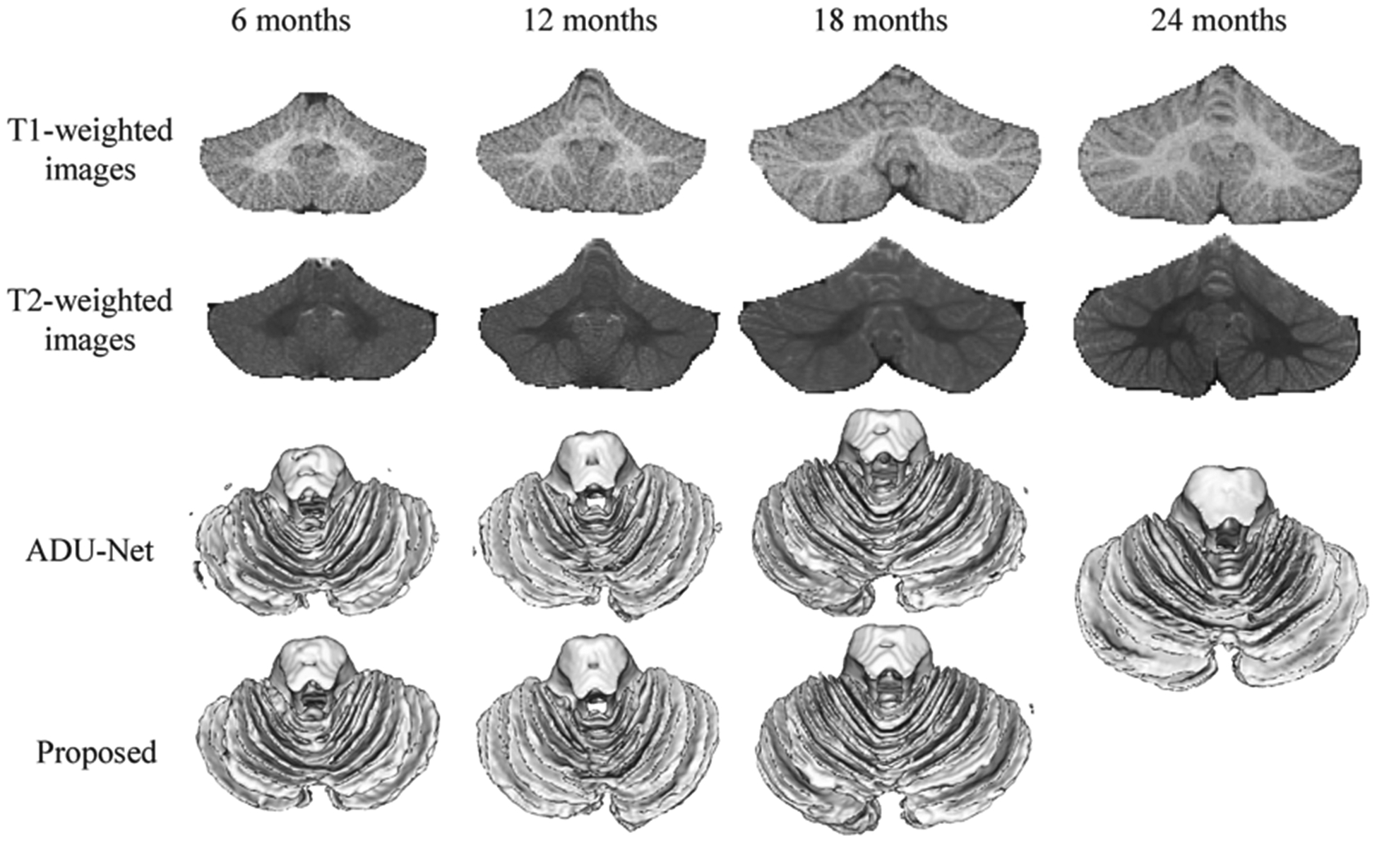
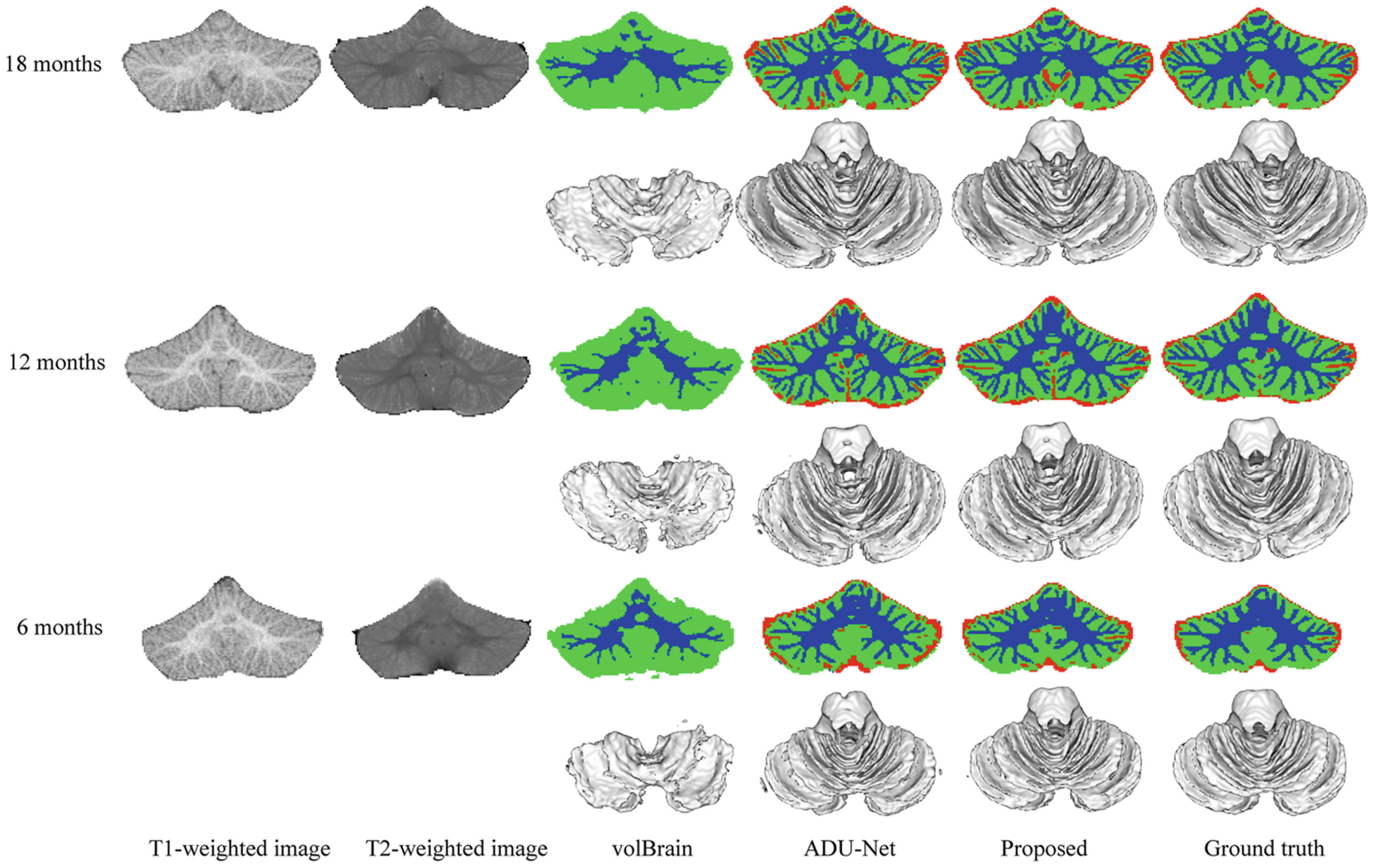
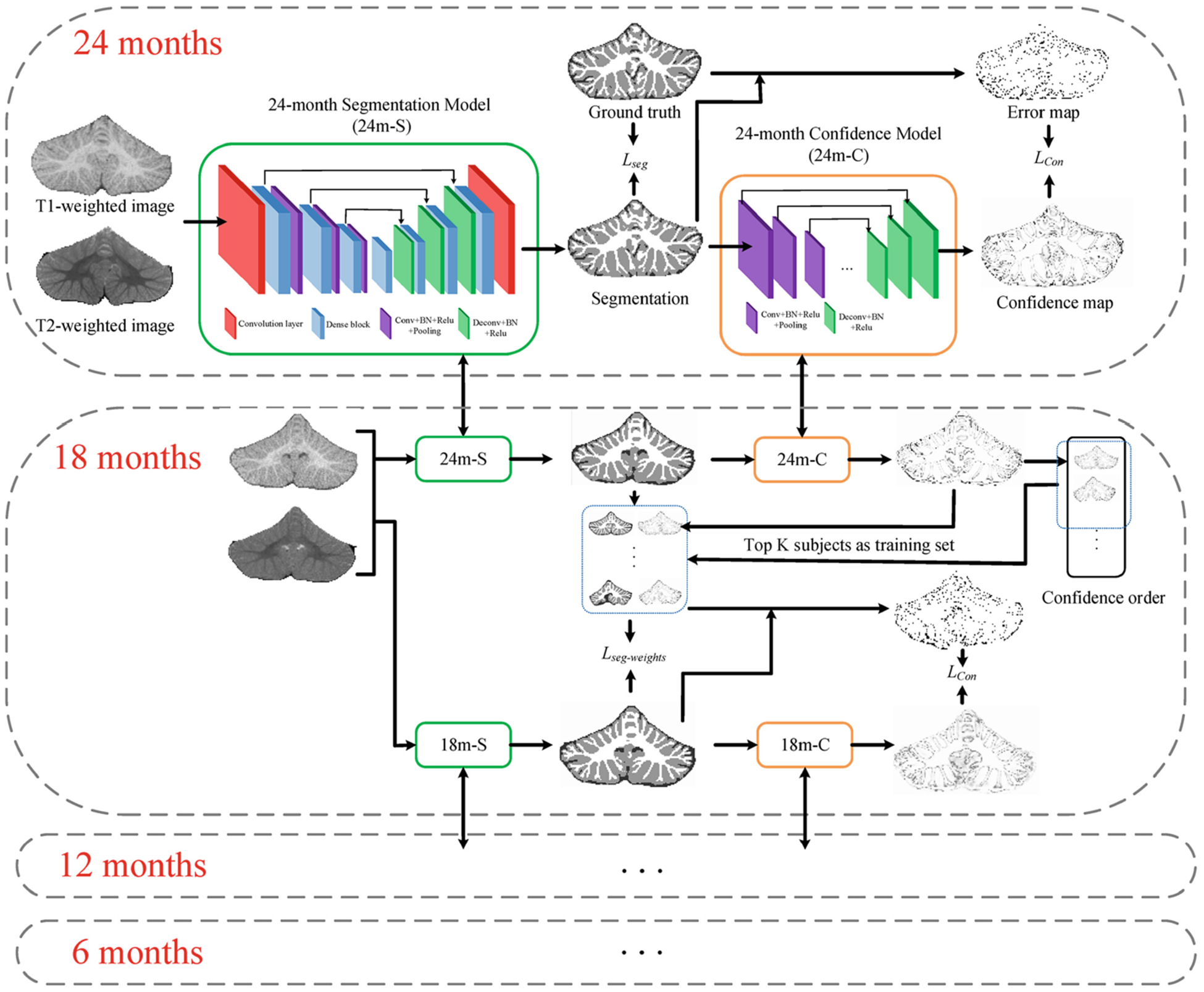
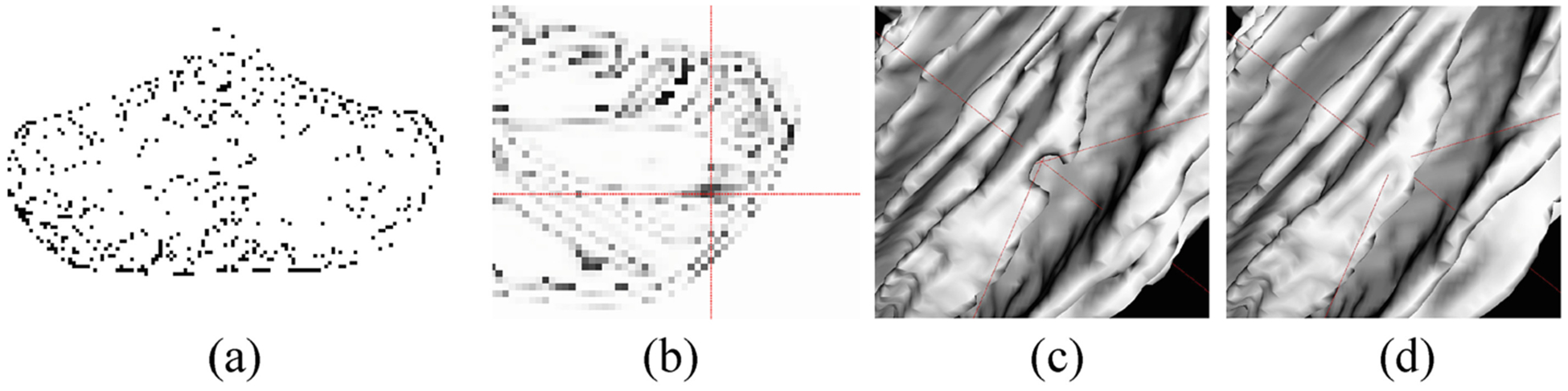
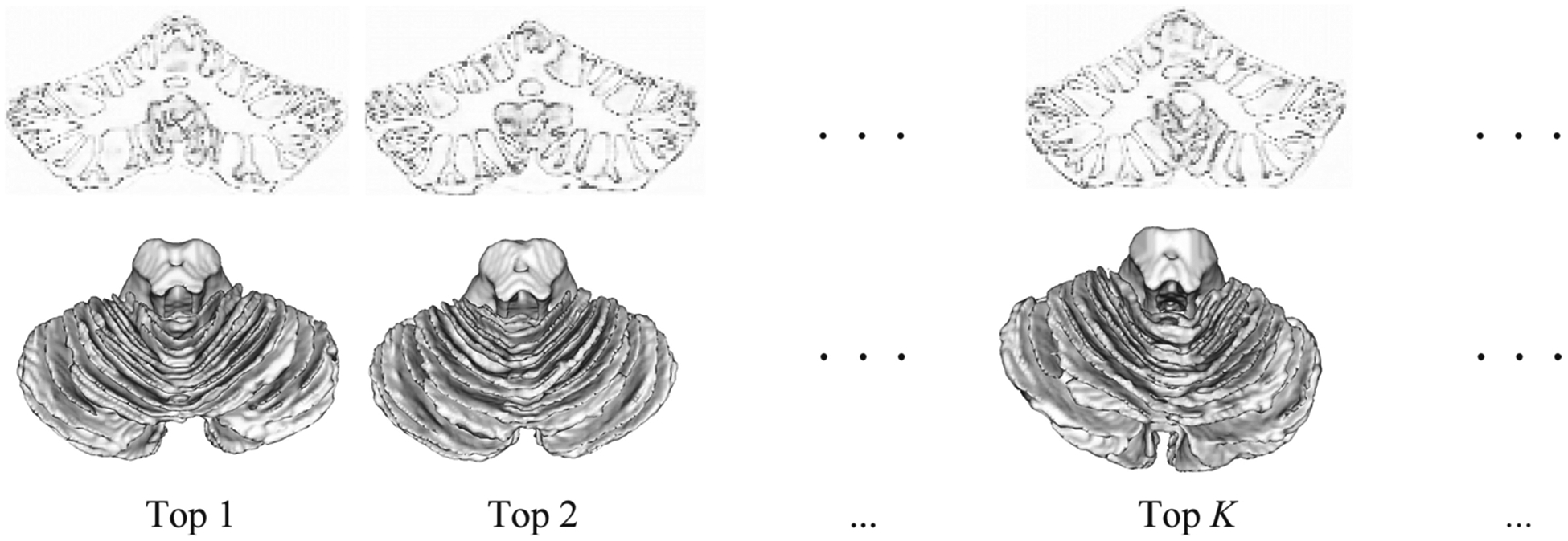
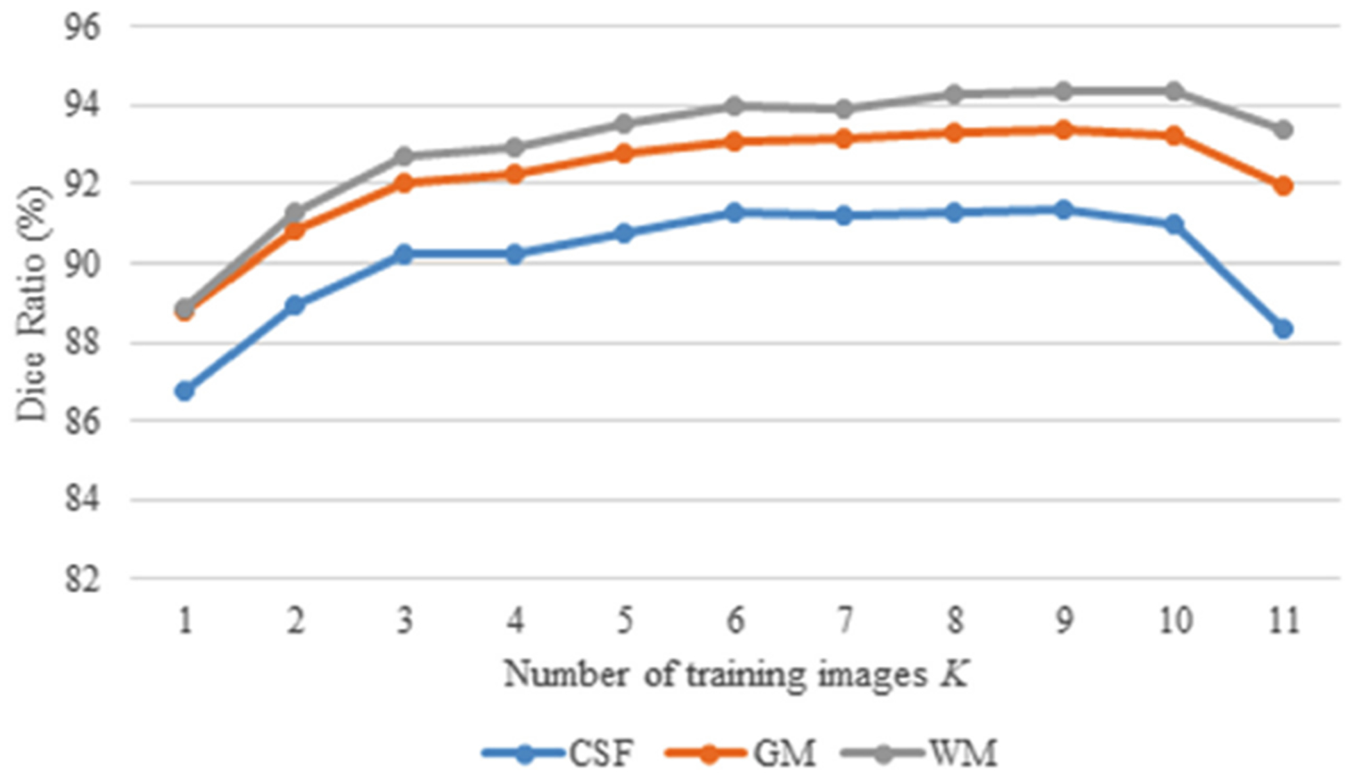
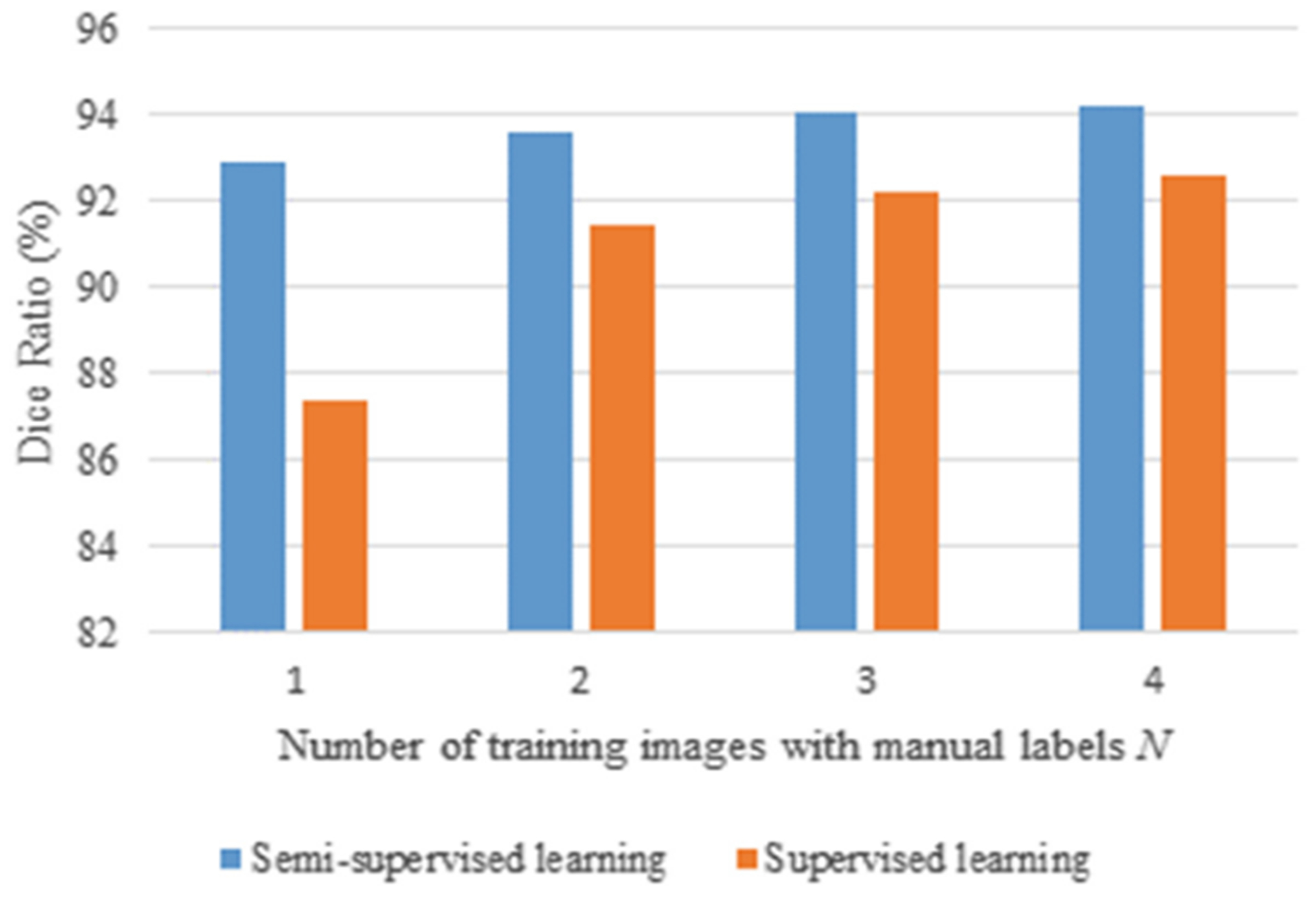
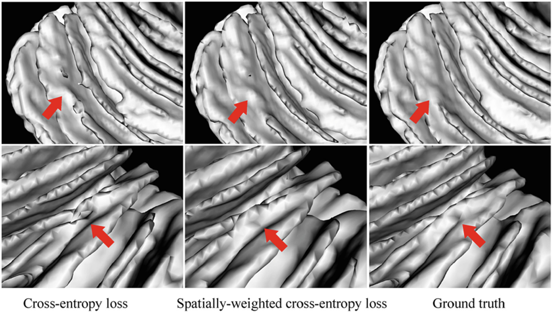
To characterize early cerebellum development, accurate segmentation of the cerebellum into white matter (WM), gray matter (GM), and cerebrospinal fluid (CSF) tissues is one of the most pivotal steps. However, due to the weak tissue contrast, extremely folded tiny structures, and severe partial volume effect, infant cerebellum tissue segmentation is especially challenging, and the manual labels are hard to obtain and correct for learning-based methods. To the best of our knowledge, there is no work on the cerebellum segmentation for infant subjects less than 24 months of age. In this work, we develop a semi-supervised transfer learning framework guided by a confidence map for tissue segmentation of cerebellum MR images from 24-month-old to 6-month-old infants. Note that only 24-month-old subjects have reliable manual labels for training, due to their high tissue contrast. Through the proposed semi-supervised transfer learning, the labels from 24-month-old subjects are gradually propagated to the 18-, 12-, and 6-month-old subjects, which have a low tissue contrast. Comparison with the state-of-the-art methods demonstrates the superior performance of the proposed method, especially for 6-month-old subjects.

摘要

为了表征小脑的早期发育,将小脑准确分割为白质(WM)、灰质(GM)和脑脊液(CSF)组织是最关键的步骤之一。然而,由于组织对比度弱、结构极度折叠且微小,以及严重的部分容积效应,婴儿小脑组织分割极具挑战性,并且手动标注难以获取,也难以用于基于学习的方法进行校正。据我们所知,尚无针对24个月以下婴儿小脑分割的研究工作。在这项研究中,我们开发了一种由置信度图引导的半监督迁移学习框架,用于对24个月至6个月大婴儿的小脑磁共振图像进行组织分割。需要注意的是,由于24个月大的受试者组织对比度高,只有他们拥有可靠的手动标注用于训练。通过所提出的半监督迁移学习,来自24个月大受试者的标注逐渐传播到组织对比度低的18个月、12个月和6个月大的受试者。与现有最先进方法的比较证明了所提方法的优越性能,特别是对于6个月大的受试者。

https://cdn.ncbi.nlm.nih.gov/pmc/blobs/740f/7885085/6fa33d46d5a1/nihms-1666988-f0008.jpg
https://cdn.ncbi.nlm.nih.gov/pmc/blobs/740f/7885085/11b0e6c807a0/nihms-1666988-f0001.jpg
https://cdn.ncbi.nlm.nih.gov/pmc/blobs/740f/7885085/3b4209006322/nihms-1666988-f0002.jpg
https://cdn.ncbi.nlm.nih.gov/pmc/blobs/740f/7885085/87974e0548a0/nihms-1666988-f0003.jpg
https://cdn.ncbi.nlm.nih.gov/pmc/blobs/740f/7885085/2a2f0324fcd1/nihms-1666988-f0004.jpg
https://cdn.ncbi.nlm.nih.gov/pmc/blobs/740f/7885085/68db3f13cd84/nihms-1666988-f0005.jpg
https://cdn.ncbi.nlm.nih.gov/pmc/blobs/740f/7885085/b32ae24a6cf2/nihms-1666988-f0006.jpg
https://cdn.ncbi.nlm.nih.gov/pmc/blobs/740f/7885085/b8ebaadb380d/nihms-1666988-f0007.jpg
https://cdn.ncbi.nlm.nih.gov/pmc/blobs/740f/7885085/6fa33d46d5a1/nihms-1666988-f0008.jpg

相似文献

1
Semi-supervised Transfer Learning for Infant Cerebellum Tissue Segmentation.用于婴儿小脑组织分割的半监督迁移学习
Mach Learn Med Imaging. 2020 Oct;12436:663-673. doi: 10.1007/978-3-030-59861-7_67. Epub 2020 Sep 29.
2
Self-supervised learning with application for infant cerebellum segmentation and analysis.基于自监督学习的婴儿小脑分割与分析。
Nat Commun. 2023 Aug 5;14(1):4717. doi: 10.1038/s41467-023-40446-z.
3
6-MONTH INFANT BRAIN MRI SEGMENTATION GUIDED BY 24-MONTH DATA USING CYCLE-CONSISTENT ADVERSARIAL NETWORKS.使用循环一致对抗网络,以24个月大的数据为指导进行6个月大婴儿脑磁共振成像分割
Proc IEEE Int Symp Biomed Imaging. 2020 Apr;2020. doi: 10.1109/isbi45749.2020.9098515. Epub 2020 May 22.
4
Automatic segmentation of MR brain images of preterm infants using supervised classification.使用监督分类对早产儿脑部磁共振图像进行自动分割。
Neuroimage. 2015 Sep;118:628-41. doi: 10.1016/j.neuroimage.2015.06.007. Epub 2015 Jun 7.
5
Learning-based 3T brain MRI segmentation with guidance from 7T MRI labeling.基于学习的3T脑磁共振成像分割,由7T磁共振成像标记引导。
Med Phys. 2016 Dec;43(12):6588-6597. doi: 10.1118/1.4967487.
6
Learning-Based 3T Brain MRI Segmentation with Guidance from 7T MRI Labeling.基于学习的3T脑磁共振成像分割与7T磁共振成像标记引导
Mach Learn Med Imaging. 2016 Oct;10019:213-220. doi: 10.1007/978-3-319-47157-0_26. Epub 2016 Oct 1.
7
Benchmark on Automatic 6-month-old Infant Brain Segmentation Algorithms: The iSeg-2017 Challenge.6个月大婴儿脑部分割算法的基准测试:iSeg-2017挑战赛
IEEE Trans Med Imaging. 2019 Feb 27. doi: 10.1109/TMI.2019.2901712.
8
Volumetric white matter tract segmentation with nested self-supervised learning using sequential pretext tasks.基于序列预训练任务的嵌套自监督学习的脑白质体积分割。
Med Image Anal. 2021 Aug;72:102094. doi: 10.1016/j.media.2021.102094. Epub 2021 Apr 30.
9
Learning-based 3T brain MRI segmentation with guidance from 7T MRI labeling.基于学习的3T脑磁共振成像分割,并以7T磁共振成像标记为指导。
Med Phys. 2016 Dec;43(12):6588. doi: 10.1118/1.4967487.
10
Automatic Accurate Infant Cerebellar Tissue Segmentation with Densely Connected Convolutional Network.基于密集连接卷积网络的自动精确婴儿小脑组织分割
Mach Learn Med Imaging. 2018 Sep;11046:233-240. doi: 10.1007/978-3-030-00919-9_27. Epub 2018 Sep 15.

引用本文的文献

1
Weakly Supervised Cerebellar Cortical Surface Parcellation with Self-Visual Representation Learning.基于自我视觉表征学习的弱监督小脑皮质表面分区
Med Image Comput Comput Assist Interv. 2023 Oct;14227:429-438. doi: 10.1007/978-3-031-43993-3_42. Epub 2023 Oct 1.
2
Deformable Spherical Transformer for Cerebellar Surface Parcellation.用于小脑表面分割的可变形球形变压器
Proc IEEE Int Symp Biomed Imaging. 2023 Apr;2023. doi: 10.1109/isbi53787.2023.10230447. Epub 2023 Sep 1.
3
Harmonized neonatal brain MR image segmentation model for cross-site datasets.

本文引用的文献

1
Multi-Site Infant Brain Segmentation Algorithms: The iSeg-2019 Challenge.多站点婴儿脑分割算法:iSeg-2019 挑战赛。
IEEE Trans Med Imaging. 2021 May;40(5):1363-1376. doi: 10.1109/TMI.2021.3055428. Epub 2021 Apr 30.
2
Developmental topography of cortical thickness during infancy.婴儿期皮质厚度的发育拓扑
Proc Natl Acad Sci U S A. 2019 Aug 6;116(32):15855-15860. doi: 10.1073/pnas.1821523116. Epub 2019 Jul 22.
3
Benchmark on Automatic 6-month-old Infant Brain Segmentation Algorithms: The iSeg-2017 Challenge.6个月大婴儿脑部分割算法的基准测试:iSeg-2017挑战赛
用于跨站点数据集的统一新生儿脑磁共振图像分割模型。
Biomed Signal Process Control. 2021 Aug;69. doi: 10.1016/j.bspc.2021.102810. Epub 2021 Jun 1.
4
Multi-Scale Self-Supervised Learning for Multi-Site Pediatric Brain MR Image Segmentation with Motion/Gibbs Artifacts.用于多站点儿科脑磁共振图像分割的多尺度自监督学习,含运动/吉布斯伪影
Mach Learn Med Imaging. 2021 Sep;12966:171-179. doi: 10.1007/978-3-030-87589-3_18. Epub 2021 Sep 21.
5
Multi-Site Infant Brain Segmentation Algorithms: The iSeg-2019 Challenge.多站点婴儿脑分割算法:iSeg-2019 挑战赛。
IEEE Trans Med Imaging. 2021 May;40(5):1363-1376. doi: 10.1109/TMI.2021.3055428. Epub 2021 Apr 30.
IEEE Trans Med Imaging. 2019 Feb 27. doi: 10.1109/TMI.2019.2901712.
4
Volume-Based Analysis of 6-Month-Old Infant Brain MRI for Autism Biomarker Identification and Early Diagnosis.基于体积分析6个月大婴儿脑部MRI以识别自闭症生物标志物并进行早期诊断。
Med Image Comput Comput Assist Interv. 2018 Sep;11072:411-419. doi: 10.1007/978-3-030-00931-1_47. Epub 2018 Sep 13.
5
Automatic Accurate Infant Cerebellar Tissue Segmentation with Densely Connected Convolutional Network.基于密集连接卷积网络的自动精确婴儿小脑组织分割
Mach Learn Med Imaging. 2018 Sep;11046:233-240. doi: 10.1007/978-3-030-00919-9_27. Epub 2018 Sep 15.
6
Exploring folding patterns of infant cerebral cortex based on multi-view curvature features: Methods and applications.基于多视图曲率特征探索婴儿大脑皮层的折叠模式:方法与应用。
Neuroimage. 2019 Jan 15;185:575-592. doi: 10.1016/j.neuroimage.2018.08.041. Epub 2018 Aug 18.
7
The UNC/UMN Baby Connectome Project (BCP): An overview of the study design and protocol development.北卡罗来纳大学教堂山分校/明尼苏达大学双城分校婴儿连接组计划(BCP):研究设计和方案制定概述。
Neuroimage. 2019 Jan 15;185:891-905. doi: 10.1016/j.neuroimage.2018.03.049. Epub 2018 Mar 22.
8
Computational neuroanatomy of baby brains: A review.婴儿大脑的计算神经解剖学:综述。
Neuroimage. 2019 Jan 15;185:906-925. doi: 10.1016/j.neuroimage.2018.03.042. Epub 2018 Mar 21.
9
Cerebellum Tissue Segmentation with Ensemble Sparse Learning.基于集成稀疏学习的小脑组织分割
Proc Int Soc Magn Reson Med Sci Meet Exhib Int Soc Magn Reson Med Sci Meet Exhib. 2017 Apr;25.
10
CERES: A new cerebellum lobule segmentation method.CERES:一种新的小脑叶分割方法。
Neuroimage. 2017 Feb 15;147:916-924. doi: 10.1016/j.neuroimage.2016.11.003. Epub 2016 Nov 8.