Marquis H, Deidda D, Gillman A, Willowson K P, Gholami Y, Hioki T, Eslick E, Thielemans K, Bailey D L
Sydney Vital Translational Cancer Research Centre, Sydney, Australia.
Institute of Medical Physics, University of Sydney, Sydney, Australia.
EJNMMI Phys. 2021 Feb 17;8(1):16. doi: 10.1186/s40658-021-00362-x.
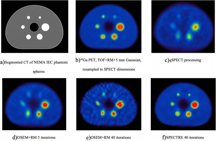
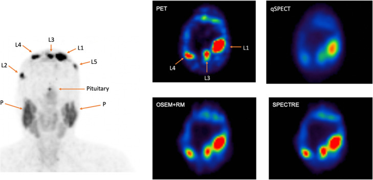
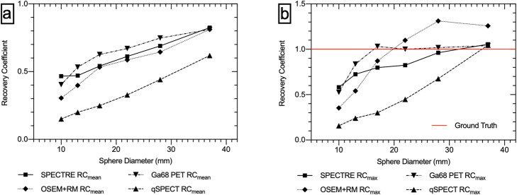
SPECT-derived dose estimates in tissues of diameter less than 3× system resolution are subject to significant losses due to the limited spatial resolution of the gamma camera. Incorporating resolution modelling (RM) into the SPECT reconstruction has been proposed as a possible solution; however, the images produced are prone to noise amplification and Gibbs artefacts. We propose a novel approach to SPECT reconstruction in a theranostic setting, which we term SPECTRE (single photon emission computed theranostic reconstruction); using a diagnostic PET image, with its superior resolution, to guide the SPECT reconstruction of the therapeutic equivalent. This report demonstrates a proof in principle of this approach.
We have employed the hybrid kernelised expectation maximisation (HKEM) algorithm implemented in STIR, with the aim of producing SPECT images with PET-equivalent resolution. We demonstrate its application in both a dual Ga/Lu IEC phantom study and a clinical example using Cu/Cu.
SPECTRE is shown to produce images comparable in accuracy and recovery to PET with minimal introduction of artefacts and amplification of noise.
The SPECTRE approach to image reconstruction shows improved quantitative accuracy with a reduction in noise amplification. SPECTRE shows great promise as a method of improving SPECT radioactivity concentrations, directly leading to more accurate dosimetry estimates in small structures and target lesions. Further investigation and optimisation of the algorithm parameters is needed before this reconstruction method can be utilised in a clinical setting.
对于直径小于系统分辨率3倍的组织,单光子发射计算机断层扫描(SPECT)得出的剂量估计值会因伽马相机有限的空间分辨率而出现显著损失。有人提出将分辨率建模(RM)纳入SPECT重建作为一种可能的解决方案;然而,生成的图像容易出现噪声放大和吉布斯伪影。我们提出了一种在诊疗一体化环境下进行SPECT重建的新方法,我们将其称为SPECTRE(单光子发射计算机诊疗重建);利用具有更高分辨率的诊断性正电子发射断层扫描(PET)图像来指导治疗等效物的SPECT重建。本报告展示了该方法的原理证明。
我们采用了在STIR中实现的混合核期望最大化(HKEM)算法,目的是生成具有PET等效分辨率的SPECT图像。我们在双镓/镥IEC体模研究和使用铜/铜的临床实例中展示了其应用。
SPECTRE所生成的图像在准确性和恢复性方面与PET相当,且伪影和噪声放大最少。
SPECTRE图像重建方法显示出更高的定量准确性,同时噪声放大有所减少。SPECTRE作为一种提高SPECT放射性浓度的方法具有很大潜力,可直接在小结构和靶病变中实现更准确的剂量测定估计。在该重建方法能够应用于临床之前,需要对算法参数进行进一步研究和优化。