Department of Radiology, Xi'an No.3 Hospital, The Affiliated Hospital of Northwest University, Shaanxi Province, 710018, Xi'an, China.
Xi'an Key Laboratory of Cardiovascular and Cerebrovascular Diseases, Xi'an No.3 Hospital, the Affiliated Hospital of Northwest University, Northwest University, Xi'an, 710018, Shaanxi Province, China.
BMC Infect Dis. 2021 Feb 18;21(1):192. doi: 10.1186/s12879-021-05839-9.
Coronavirus disease 2019 (COVID-19) has caused a global pandemic that has raised worldwide concern. This study aims to investigate the correlation between the extent of lung infection and relevant clinical laboratory testing indicators in COVID-19 and to analyse its underlying mechanism.
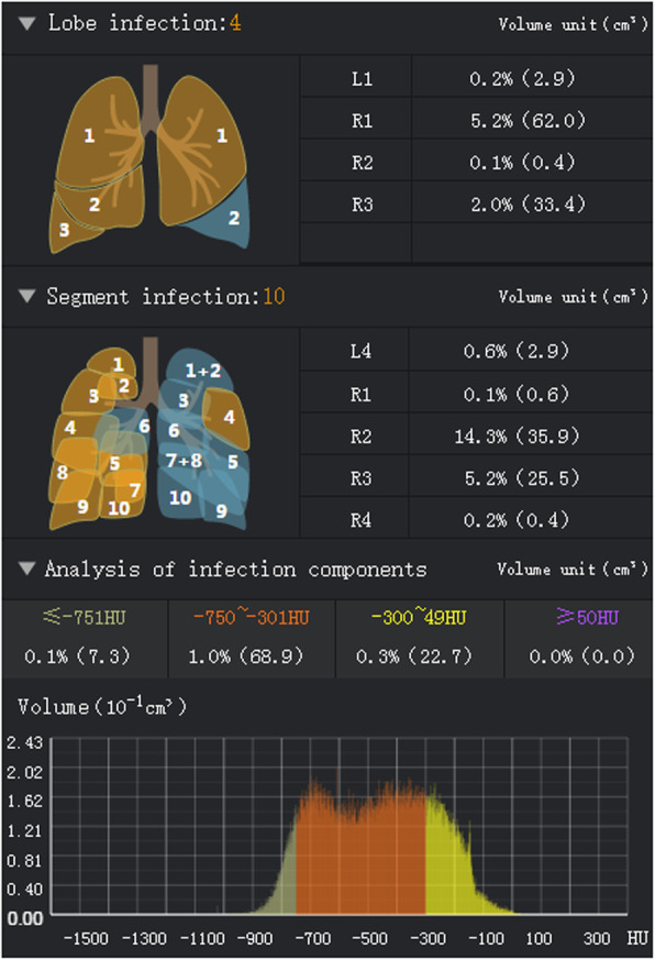
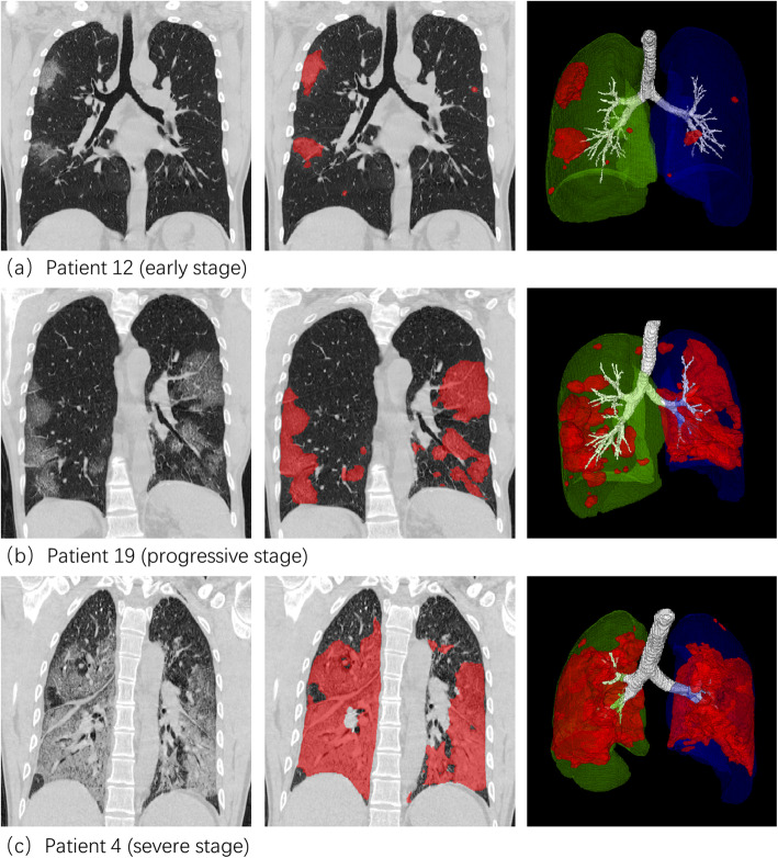
Chest high-resolution computer tomography (CT) images and laboratory examination data of 31 patients with COVID-19 were extracted, and the lesion areas in CT images were quantitatively segmented and calculated using a deep learning (DL) system. A cross-sectional study method was carried out to explore the differences among the proportions of lung lobe infection and to correlate the percentage of infection (POI) of the whole lung in all patients with clinical laboratory examination values.
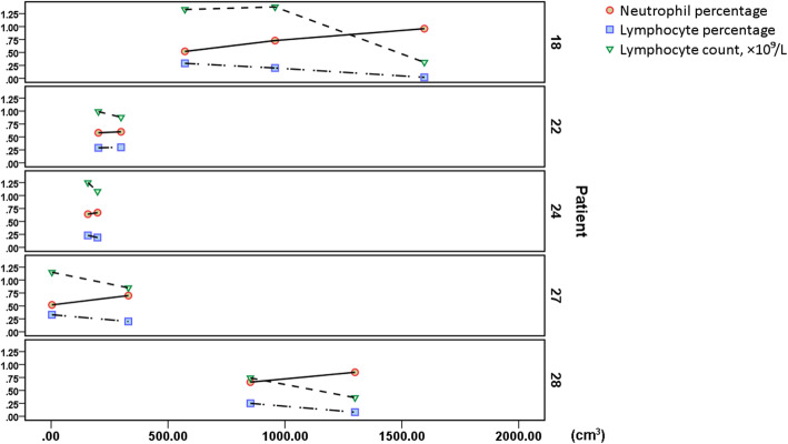
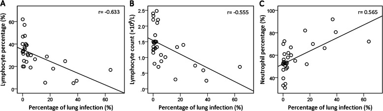
No significant difference in the proportion of infection was noted among various lung lobes (P > 0.05). The POI of total lung was negatively correlated with the peripheral blood lymphocyte percentage (L%) (r = - 0.633, P < 0.001) and lymphocyte (LY) count (r = - 0.555, P = 0.001) but positively correlated with the neutrophil percentage (N%) (r = 0.565, P = 0.001). Otherwise, the POI was not significantly correlated with the peripheral blood white blood cell (WBC) count, monocyte percentage (M%) or haemoglobin (HGB) content. In some patients, as the infection progressed, the L% and LY count decreased progressively accompanied by a continuous increase in the N%.
Lung lesions in COVID-19 patients are significantly correlated with the peripheral blood lymphocyte and neutrophil levels, both of which could serve as prognostic indicators that provide warning implications, and contribute to clinical interventions in patients.
2019 年冠状病毒病(COVID-19)已在全球范围内引起关注。本研究旨在探讨 COVID-19 中肺部感染程度与相关临床实验室检测指标的相关性,并分析其潜在机制。
提取了 31 例 COVID-19 患者的胸部高分辨率计算机断层扫描(CT)图像和实验室检查数据,使用深度学习(DL)系统对 CT 图像中的病变区域进行定量分割和计算。采用横断面研究方法,探讨各肺叶感染比例的差异,并将所有患者的全肺感染百分比(POI)与临床实验室检查值进行相关性分析。
各肺叶的感染比例无显著差异(P>0.05)。全肺 POI 与外周血淋巴细胞百分比(L%)(r=-0.633,P<0.001)和淋巴细胞(LY)计数(r=-0.555,P=0.001)呈负相关,与中性粒细胞百分比(N%)(r=0.565,P=0.001)呈正相关。然而,POI 与外周血白细胞(WBC)计数、单核细胞百分比(M%)或血红蛋白(HGB)含量无显著相关性。在一些患者中,随着感染的进展,L%和 LY 计数逐渐降低,而 N%持续增加。
COVID-19 患者的肺部病变与外周血淋巴细胞和中性粒细胞水平显著相关,这两者都可以作为预后指标,为临床干预提供预警作用。