Liu Jieke, Xu Hao, Qing Haomiao, Li Yong, Yang Xi, He Changjiu, Ren Jing, Zhou Peng
Department of Radiology, Sichuan Cancer Hospital & Institute, Sichuan Cancer Center, School of Medicine, University of Electronic Science and Technology of China, Chengdu, China.
Front Oncol. 2021 Feb 2;10:634298. doi: 10.3389/fonc.2020.634298. eCollection 2020.
This study aimed to develop radiomic models based on low-dose CT (LDCT) and standard-dose CT to distinguish adenocarcinomas from benign lesions in patients with solid solitary pulmonary nodules and compare the performance among these radiomic models and Lung CT Screening Reporting and Data System (Lung-RADS). The reproducibility of radiomic features between LDCT and standard-dose CT were also evaluated.
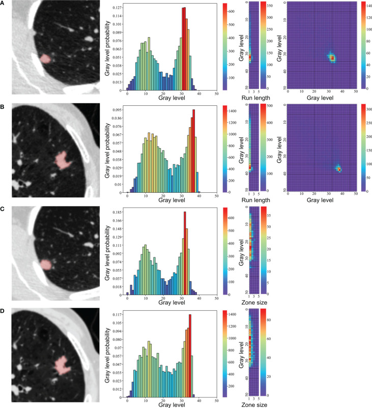
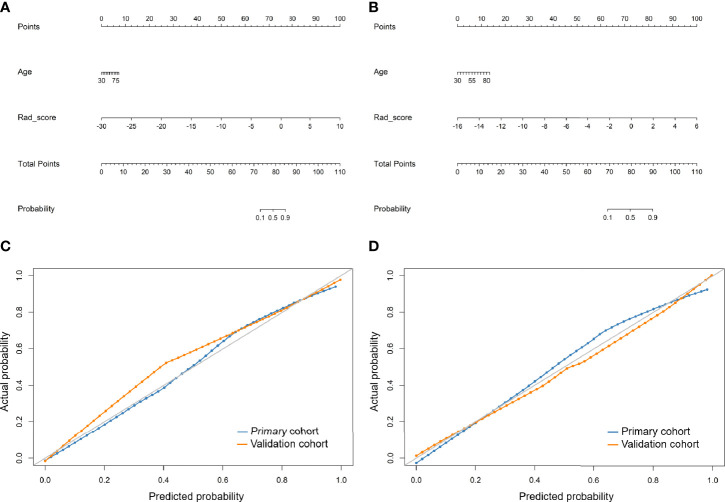
A total of 141 consecutive pathologically confirmed solid solitary pulmonary nodules were enrolled including 50 adenocarcinomas and 48 benign nodules in primary cohort and 22 adenocarcinomas and 21 benign nodules in validation cohort. LDCT and standard-dose CT scans were conducted using same acquisition parameters and reconstruction method except for radiation dose. All nodules were automatically segmented and 104 original radiomic features were extracted. The concordance correlation coefficient was used to quantify reproducibility of radiomic features between LDCT and standard-dose CT. Radiomic features were selected to build radiomic signature, and clinical characteristics and radiomic signature were combined to develop radiomic nomogram for LDCT and standard-dose CT, respectively. The performance of radiomic models and Lung-RADS was assessed by area under curve (AUC) of receiver operating characteristic curve, sensitivity, and specificity.
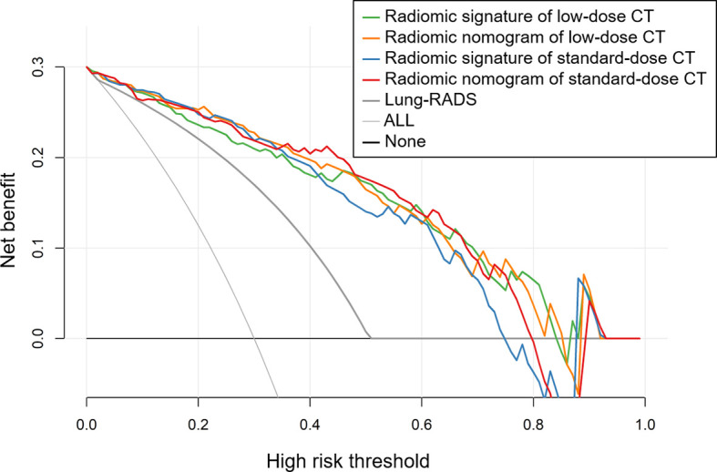
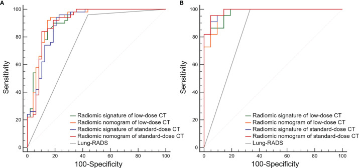
Shape and first order features, and neighboring gray tone difference matrix features were highly reproducible between LDCT and standard-dose CT. No significant differences of AUCs were found among radiomic signature and nomogram of LDCT and standard-dose CT in both primary and validation cohort (0.915 0.919 0.898 0.909 and 0.976 0.976 0.985 0.987, respectively). These radiomic models had higher specificity than Lung-RADS (all correct < 0.05), while there were no significant differences of sensitivity between Lung-RADS and radiomic models.
The diagnostic performance of LDCT-based radiomic models to differentiate adenocarcinomas from benign lesions in solid pulmonary nodules were equivalent to that of standard-dose CT. The LDCT-based radiomic model with higher specificity and lower false-positive rate than Lung-RADS might help reduce overdiagnosis and overtreatment of solid pulmonary nodules in lung cancer screening.
本研究旨在基于低剂量CT(LDCT)和标准剂量CT开发放射组学模型,以区分实性孤立性肺结节患者的腺癌与良性病变,并比较这些放射组学模型与肺部CT筛查报告和数据系统(Lung-RADS)的性能。还评估了LDCT和标准剂量CT之间放射组学特征的可重复性。
共纳入141例经病理证实的连续实性孤立性肺结节,其中原发性队列中有50例腺癌和48例良性结节,验证队列中有22例腺癌和21例良性结节。除辐射剂量外,LDCT和标准剂量CT扫描采用相同的采集参数和重建方法。所有结节均自动分割,并提取104个原始放射组学特征。采用一致性相关系数量化LDCT和标准剂量CT之间放射组学特征的可重复性。选择放射组学特征构建放射组学特征图谱,并分别结合临床特征和放射组学特征图谱开发LDCT和标准剂量CT的放射组学列线图。通过受试者操作特征曲线的曲线下面积(AUC)、敏感性和特异性评估放射组学模型和Lung-RADS的性能。
LDCT和标准剂量CT之间的形状和一阶特征以及邻域灰度差矩阵特征具有高度可重复性。在原发性队列和验证队列中,LDCT和标准剂量CT的放射组学特征图谱和列线图的AUCs均无显著差异(分别为0.915、0.919、0.898、0.909和0.976、0.976、0.985、0.987)。这些放射组学模型的特异性高于Lung-RADS(所有P<0.05),而Lung-RADS和放射组学模型之间的敏感性无显著差异。
基于LDCT的放射组学模型区分实性肺结节中腺癌与良性病变的诊断性能与标准剂量CT相当。基于LDCT的放射组学模型比Lung-RADS具有更高的特异性和更低的假阳性率,可能有助于减少肺癌筛查中实性肺结节的过度诊断和过度治疗。