Centre for Atherothrombosis and Metabolic Disease, Hull York Medical School, University of Hull, Hull, United Kingdom.
Diabetes, Endocrinology and Metabolism, Hull York Medical School, University of Hull, Hull, United Kingdom.
PLoS One. 2021 Feb 19;16(2):e0247234. doi: 10.1371/journal.pone.0247234. eCollection 2021.
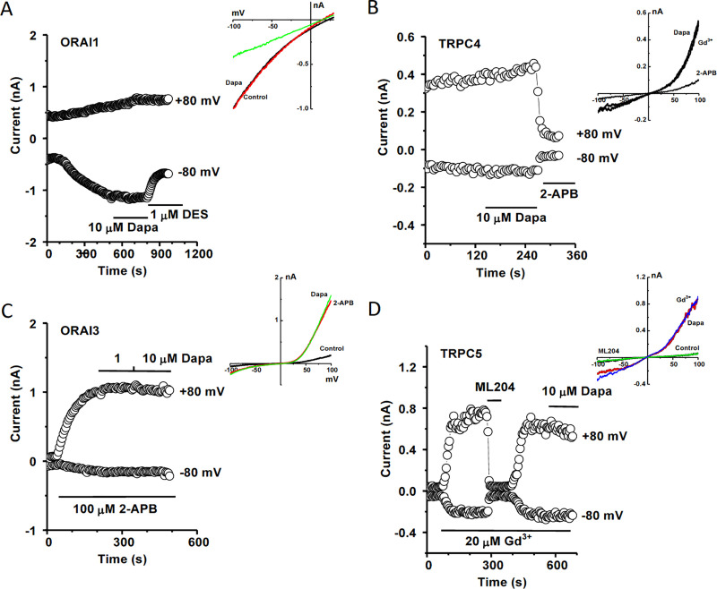
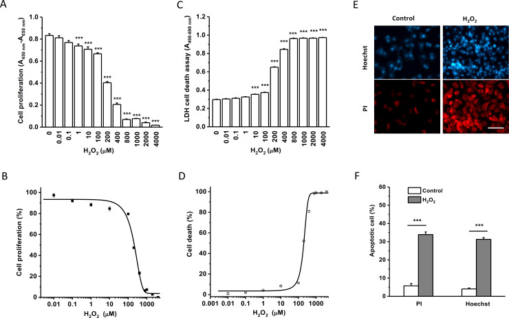
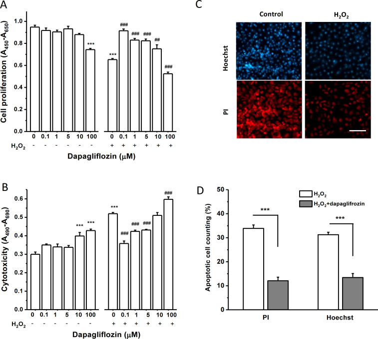
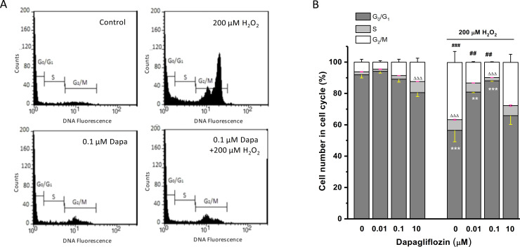
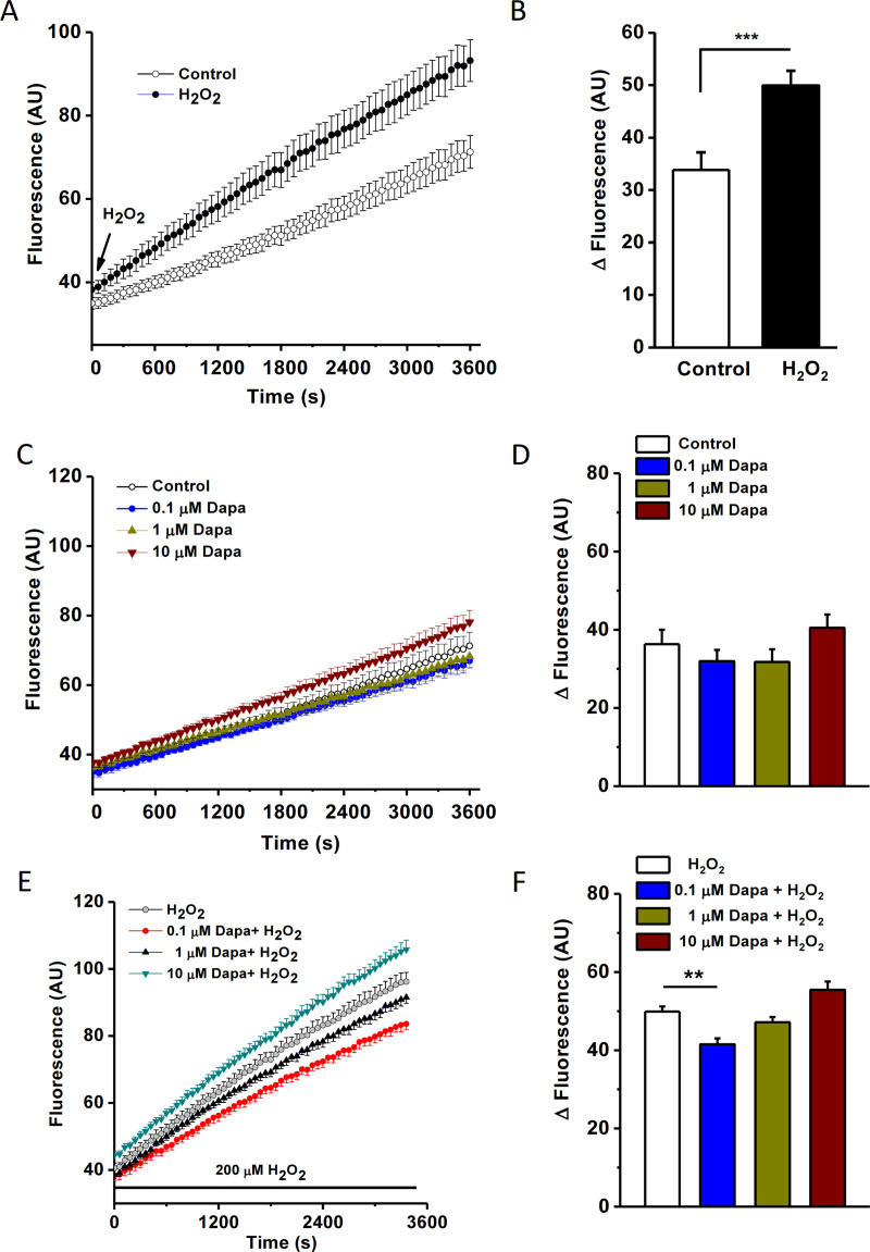
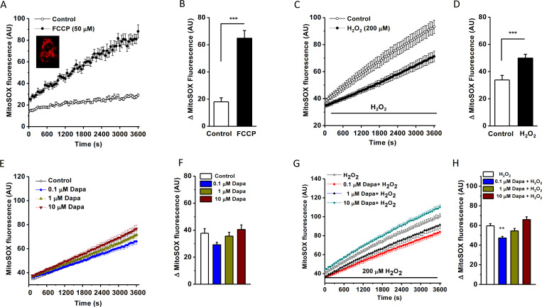
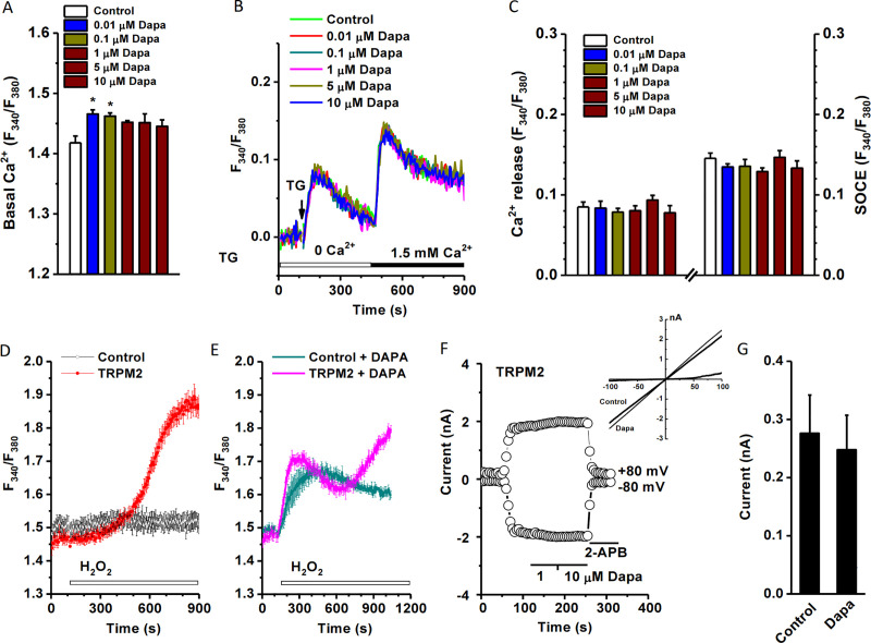
Elevated reactive oxygen species (ROS) in type 2 diabetes cause cellular damage in many organs. Recently, the new class of glucose-lowering agents, SGLT-2 inhibitors, have been shown to reduce the risk of developing diabetic complications; however, the mechanisms of such beneficial effect are largely unknown. Here we aimed to investigate the effects of dapagliflozin on cell proliferation and cell death under oxidative stress conditions and explore its underlying mechanisms. Human proximal tubular cells (HK-2) were used. Cell growth and death were monitored by cell counting, water-soluble tetrazolium-1 (WST-1) and lactate dehydrogenase (LDH) assays, and flow cytometry. The cytosolic and mitochondrial (ROS) production was measured using fluorescent probes (H2DCFDA and MitoSOX) under normal and oxidative stress conditions mimicked by addition of H2O2. Intracellular Ca2+ dynamics was monitored by FlexStation 3 using cell-permeable Ca2+ dye Fura-PE3/AM. Dapagliflozin (0.1-10 μM) had no effect on HK-2 cell proliferation under normal conditions, but an inhibitory effect was seen at an extreme high concentration (100 μM). However, dapagliflozin at 0.1 to 5 μM showed remarkable protective effects against H2O2-induced cell injury via increasing the viable cell number at phase G0/G1. The elevated cytosolic and mitochondrial ROS under oxidative stress was significantly decreased by dapagliflozin. Dapagliflozin increased the basal intracellular [Ca2+]i in proximal tubular cells, but did not affect calcium release from endoplasmic reticulum and store-operated Ca2+ entry. The H2O2-sensitive TRPM2 channel seemed to be involved in the Ca2+ dynamics regulated by dapagliflozin. However, dapagliflozin had no direct effects on ORAI1, ORAI3, TRPC4 and TRPC5 channels. Our results suggest that dapagliflozin shows anti-oxidative properties by reducing cytosolic and mitochondrial ROS production and altering Ca2+ dynamics, and thus exerts its protective effects against cell damage under oxidative stress environment.
在 2 型糖尿病中,活性氧(ROS)的升高会导致许多器官的细胞损伤。最近,一类新型的降血糖药物 SGLT-2 抑制剂已被证明可降低发生糖尿病并发症的风险;然而,这种有益作用的机制在很大程度上尚不清楚。在这里,我们旨在研究达格列净在氧化应激条件下对细胞增殖和细胞死亡的影响,并探讨其潜在机制。使用人近端肾小管细胞(HK-2)进行研究。通过细胞计数、水溶性四唑盐-1(WST-1)和乳酸脱氢酶(LDH)测定以及流式细胞术监测细胞生长和死亡。在正常条件下以及通过添加 H2O2 模拟的氧化应激条件下,使用荧光探针(H2DCFDA 和 MitoSOX)测量细胞质和线粒体(ROS)的产生。通过 FlexStation 3 使用细胞通透性 Ca2+染料 Fura-PE3/AM 监测细胞内 Ca2+动力学。达格列净(0.1-10 μM)在正常条件下对 HK-2 细胞增殖没有影响,但在极高浓度(100 μM)下观察到抑制作用。然而,达格列净在 0.1 至 5 μM 浓度下表现出对 H2O2 诱导的细胞损伤的显著保护作用,通过增加 G0/G1 期的存活细胞数量来实现。氧化应激下升高的细胞质和线粒体 ROS 水平显著被达格列净降低。达格列净增加了近端肾小管细胞的基础细胞内 [Ca2+]i,但不影响内质网钙释放和储存操纵的 Ca2+内流。H2O2 敏感的 TRPM2 通道似乎参与了达格列净调节的 Ca2+动力学。然而,达格列净对 ORAI1、ORAI3、TRPC4 和 TRPC5 通道没有直接作用。我们的研究结果表明,达格列净通过降低细胞质和线粒体 ROS 的产生和改变 Ca2+动力学表现出抗氧化特性,并因此在氧化应激环境下发挥对细胞损伤的保护作用。