• 文献检索
  • 文档翻译
  • 深度研究
  • 学术资讯
  • Suppr Zotero 插件Zotero 插件
  • 邀请有礼
  • 套餐&价格
  • 历史记录
应用&插件
Suppr Zotero 插件Zotero 插件浏览器插件Mac 客户端Windows 客户端微信小程序
定价
高级版会员购买积分包购买API积分包
服务
文献检索文档翻译深度研究API 文档MCP 服务
关于我们
关于 Suppr公司介绍联系我们用户协议隐私条款
关注我们

Suppr 超能文献

核心技术专利:CN118964589B侵权必究
粤ICP备2023148730 号-1Suppr @ 2026

文献检索

告别复杂PubMed语法,用中文像聊天一样搜索,搜遍4000万医学文献。AI智能推荐,让科研检索更轻松。

立即免费搜索

文件翻译

保留排版,准确专业,支持PDF/Word/PPT等文件格式,支持 12+语言互译。

免费翻译文档

深度研究

AI帮你快速写综述,25分钟生成高质量综述,智能提取关键信息,辅助科研写作。

立即免费体验

三种牙科手术显微镜光学性能的比较:一项初步研究。

Comparison of optical performance among three dental operating microscopes: A pilot study.

作者信息

Jalali Poorya, Kim Charles, Woodmansey Karl F

机构信息

Departments of Endodontics, Texas A and M College of Dentistry, Dallas, Texas, USA.

出版信息

J Conserv Dent. 2020 Jul-Aug;23(4):374-376. doi: 10.4103/JCD.JCD_191_20. Epub 2021 Jan 16.

DOI:10.4103/JCD.JCD_191_20
PMID:33623239
原文链接:https://pmc.ncbi.nlm.nih.gov/articles/PMC7883792/
Abstract

INTRODUCTION

Two important aspects of the dental operating microscope (DOM) that factor into its overall effectiveness are resolution and depth of field. Therefore, the objective of this study was to evaluate and compare the resolution and depth of field of DOMs from three well-known manufacturers using standardized test targets.

MATERIALS AND METHODS

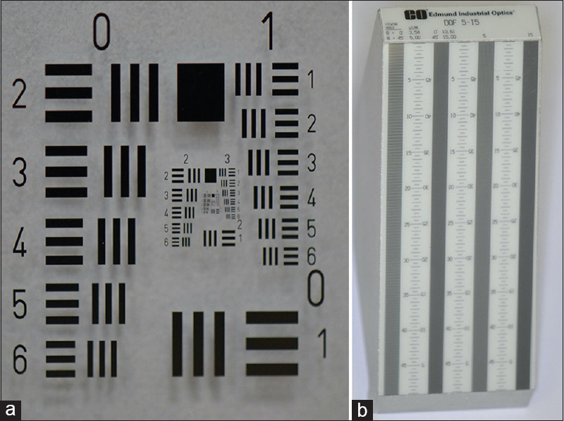
A resolution test, using the 1951 USAF Hi-Resolution Target (Edmund Optics, Barrington, NJ), and a depth of field test, using the Depth of Field Target 5-15 (Edmund Optics, Barrington, NJ), were performed by two calibrated observers. Three DOM systems such as Seiler IQ (Seiler Instrument Inc., St. Louis, USA), Global G-Series 6 step (Global Surgical Corp., St. Louis, USA), and Zeiss Extaro 300 (Carl Zeiss Meditec AG, Oberkochen, Germany) were used to compare the resolution and depth of field.

RESULTS

The Zeiss Extaro 300 showed the highest maximum resolution and maximum DOF (64 lp/mm and 17mm, respectively). The Seiler IQ showed the lowest maximum resolution and maximum DOF (35.9 lp/mm and 11 mm, respectively).

CONCLUSION

Within the limitations of this study, the Zeiss Extaro 300 was superior in terms of resolution and depth of field as compared to the other two DOMs.

摘要

引言

牙科手术显微镜(DOM)整体效能的两个重要因素是分辨率和景深。因此,本研究的目的是使用标准化测试靶标评估和比较三个知名制造商生产的DOM的分辨率和景深。

材料与方法

由两名经过校准的观察者进行分辨率测试,使用1951年美国空军高分辨率靶标(埃德蒙光学公司,新泽西州巴林顿),以及景深测试,使用景深靶标5-15(埃德蒙光学公司,新泽西州巴林顿)。使用三种DOM系统,如Seiler IQ(美国密苏里州圣路易斯市Seiler仪器公司)、Global G-Series 6 step(美国密苏里州圣路易斯市Global Surgical Corp.)和蔡司Extaro 300(德国奥伯科亨市卡尔蔡司医疗技术股份公司)来比较分辨率和景深。

结果

蔡司Extaro 300显示出最高的最大分辨率和最大景深(分别为64线对/毫米和17毫米)。Seiler IQ显示出最低的最大分辨率和最大景深(分别为35.9线对/毫米和11毫米)。

结论

在本研究的局限性内,与其他两种DOM相比,蔡司Extaro 300在分辨率和景深方面更具优势。

https://cdn.ncbi.nlm.nih.gov/pmc/blobs/888f/7883792/5e1671744d26/JCD-23-374-g001.jpg

相似文献

1
Comparison of optical performance among three dental operating microscopes: A pilot study.三种牙科手术显微镜光学性能的比较:一项初步研究。
J Conserv Dent. 2020 Jul-Aug;23(4):374-376. doi: 10.4103/JCD.JCD_191_20. Epub 2021 Jan 16.
2
Comparison of a Dental Operating Microscope and High-resolution Videoscope for Endodontic Procedures.口腔手术显微镜与高分辨率内镜在根管治疗术中的比较。
J Endod. 2020 May;46(5):688-693. doi: 10.1016/j.joen.2020.01.013. Epub 2020 Mar 2.
3
[The Development of First Operating Microscopes from Zeiss Jena and Oberkochen to Series Production].[从蔡司耶拿和奥伯科亨的初代手术显微镜到批量生产的发展历程]
Laryngorhinootologie. 2017 Jan;96(1):27-34. doi: 10.1055/s-0042-104416. Epub 2016 Apr 29.
4
Subjective and objective depth of field measures in pseudophakic eyes: comparison between extended depth of focus, trifocal and bifocal intraocular lenses.人工晶状体眼的主观和客观景深测量:扩展景深、三焦点和双焦点人工晶状体的比较。
Int Ophthalmol. 2020 Feb;40(2):351-359. doi: 10.1007/s10792-019-01186-6. Epub 2019 Oct 3.
5
Single-Surgeon in Vivo Experience with the Zeiss QEVO Microinspection Tool: An Analysis of Its Use for Extending the Reach of Operative Visualization.蔡司 QEVO 微检测工具的单外科医生活体经验:分析其用于扩展手术可视化范围的应用。
World Neurosurg. 2021 Mar;147:268-272. doi: 10.1016/j.wneu.2020.12.036.
6
New method for determining the depth of field of microscope systems.确定显微镜系统景深的新方法。
Appl Opt. 2011 Oct 1;50(28):5524-33. doi: 10.1364/AO.50.005524.
7
Laboratory evaluation of the optical properties of two extended-depth-of-focus intraocular lenses.两种扩展景深人工晶状体光学特性的实验室评估。
BMC Ophthalmol. 2020 Feb 14;20(1):53. doi: 10.1186/s12886-020-1332-6.
8
Combining optical coherence tomography with visual field data to rapidly detect disease progression in glaucoma: a diagnostic accuracy study.结合光学相干断层扫描和视野数据快速检测青光眼疾病进展:一项诊断准确性研究。
Health Technol Assess. 2018 Jan;22(4):1-106. doi: 10.3310/hta22040.
9
[Back-calculation of the keratometer index-Which value would have been correct in cataract surgery?].[角膜曲率计指数的反推计算——白内障手术中哪个值才是正确的?]
Ophthalmologe. 2021 Apr;118(4):356-366. doi: 10.1007/s00347-020-01182-7.
10
Comparison of optics and performance of a distal sensor high definition cystoscope, a distal sensor standard definition cystoscope, and a fiberoptic cystoscope.比较远端传感器高清膀胱镜、远端传感器标准定义膀胱镜和纤维膀胱镜的光学性能和性能。
Urology. 2013 Dec;82(6):1226-30. doi: 10.1016/j.urology.2013.06.033. Epub 2013 Oct 2.

引用本文的文献

1
Depth of field or depth of focus?景深还是焦深?
J Cataract Refract Surg. 2024 Dec 1;50(12):1291-1292. doi: 10.1097/j.jcrs.0000000000001548.
2
Platelet-rich fibrin as an apical barrier for MTA placement in the treatment of teeth with open apices: a pilot study.富血小板纤维蛋白作为在根尖敞开的牙齿中放置 MTA 的顶端屏障的疗效:一项初步研究。
J Transl Med. 2024 May 25;22(1):498. doi: 10.1186/s12967-024-05318-0.

本文引用的文献

1
Comparison of a Dental Operating Microscope and High-resolution Videoscope for Endodontic Procedures.口腔手术显微镜与高分辨率内镜在根管治疗术中的比较。
J Endod. 2020 May;46(5):688-693. doi: 10.1016/j.joen.2020.01.013. Epub 2020 Mar 2.
2
Magnification in endodontics: A review of its application and acceptance among dental practitioners.牙髓病学中的放大技术:牙科从业者对其应用及接受情况的综述
Eur J Dent. 2018 Oct-Dec;12(4):610-616. doi: 10.4103/ejd.ejd_248_18.
3
Influence of operating microscope in the sealing of cervical perforations.
手术显微镜在封闭宫颈穿孔中的作用
J Conserv Dent. 2016 Mar-Apr;19(2):152-6. doi: 10.4103/0972-0707.178695.
4
Magnification alternatives: seeing is believing, Part I.
Dent Today. 2013 Jun;32(6):82-7.
5
Non surgical perforation repair by mineral trioxide aggregate under dental operating microscope.牙科手术显微镜下用三氧化矿物凝聚体进行非手术性穿孔修复
J Conserv Dent. 2011 Jan;14(1):83-5. doi: 10.4103/0972-0707.80729.
6
Magnification's effect on endodontic fine motor skills.放大倍数对牙髓精细运动技能的影响。
J Endod. 2010 Jul;36(7):1135-8. doi: 10.1016/j.joen.2010.03.003. Epub 2010 Apr 18.
7
The use of the operating microscope in endodontics.手术显微镜在牙髓病学中的应用。
Dent Clin North Am. 2010 Apr;54(2):191-214. doi: 10.1016/j.cden.2010.01.002.
8
Canal morphology of maxillary molars: clinical observations of canal configurations.上颌磨牙的根管形态:根管形态的临床观察
J Endod. 1999 Jun;25(6):446-50. doi: 10.1016/S0099-2399(99)80276-3.
9
Periodontal microsurgery.牙周显微外科手术
Dent Clin North Am. 1998 Apr;42(2):339-59.
10
Microscopes in endodontics.牙髓病学中的显微镜
J Calif Dent Assoc. 1992 Nov;20(11):55-61.