Liu Lei, Mo Yanfei, Wu Bingying, Yu Zongliang, Sun Bugao
Department of Cardiology, Affiliated Drum Tower Hospital of Nanjing University, Nanjing, China.
Department of Cardiology, Nanjing Pukou Hospital of TCM, Nanjing, China.
Evid Based Complement Alternat Med. 2020 Dec 3;2020:8762509. doi: 10.1155/2020/8762509. eCollection 2020.
Poge heart-saving decoction (PHSD) has been used as a medicine treating heart failure in China for many years. The study aimed to explore the effect of PHSD on cardiac function in heart failure conditions and its underlying mechanism.
Adriamycin was used to induce the model of heart failure (HF) in rats. Sixty rats were randomly divided into six groups: blank control group, sham group, 9.33 g/kg group (low-PHSD, test group), 13.995 g/kg group (moderate-PHSD, test group), 18.66 g/kg group (high-PHSD, test group), and fosinopril group (4.67 mg/kg, comparison test group). Cardiac ultrasound was used to evaluate the cardiac function of the rats, and radioimmunoassay was used to measure aldosterone (ALD) and angiotensin II (AngII) levels in the serum.
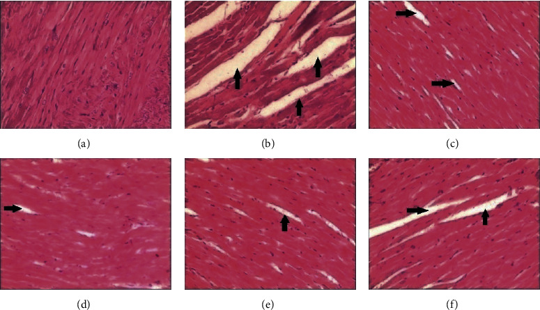
Compared with the blank control group, the left ventricular end-diastolic dimension (LVEDd) and left ventricular end-systolic dimension (LVEDs) in the sham group were increased (1.04 ± 0.12 vs. 0.67 ± 0.13 cm; 0.75 ± 0.13 vs. 0.28 ± 0.10 cm; < 0.05), and the left ventricular ejection fraction was decreased (36.65 ± 5.74 vs. 76.09 ± 4.23%; < 0.05). The ejection fraction of HF rats was increased in 9.33 g/kg group, 13.995 g/kg group, and 18.66 g/kg group compared with those of the sham group (57.13 ± 1.63, 58.43 ± 1.98, and 59.21 ± 1.37 vs. 36.65 ± 5.74%; < 0.05). PHSD also improved cardiac function by reducing the LVEDd and LVEDs (0.88 ± 0.11, 0.75 ± 0.13, and 0.72 ± 0.18 vs. 1.04 ± 0.12 cm; 0.62 ± 0.10, 0.63 ± 0.17, and 0.45 ± 0.11 vs. 0.75 ± 0.13 cm; < 0.05). The levels of ALD and AngII in the serum of rats in the sham group were significantly higher than those in the blank control group (371.58 ± 39.25 vs. 237.12 ± 17.35 g/L; 232.18 ± 16.33 vs. 159.44 ± 18.42 pg/L; < 0.05). The ALD and AngII of the rats in all of the three PHSD groups and the fosinopril group were decreased (276.81 ± 25.63, 277.18 ± 21.35, 268.19 ± 19.28, and 271.47 ± 28.96 vs. 371.58 ± 39.25 g/L; 169.41 ± 27.53, 168.81 ± 19.78, 164.23 ± 21.34, and 174.27 ± 22.84 vs. 232.18 ± 16.33 pg/L; < 0.05). The histopathological changes of the myocardium in the sham group showed the disorganized fiber, shaded staining, fracture, and zonation. The fracture of the myocardium was relieved in all groups except the sham group and the blank control group.
Therefore, PHSD could shorten LVEDd and LVEDs of rats and reverse ventricular remodeling. The mechanism might be related to the inhibition of the activation level of renin-angiotensin-aldosterone system (especially ALD and AngII) and decreasing the postload of the heart.
芪苈强心汤在中国作为治疗心力衰竭的药物已应用多年。本研究旨在探讨芪苈强心汤对心力衰竭大鼠心功能的影响及其潜在机制。
采用阿霉素诱导大鼠心力衰竭模型。60只大鼠随机分为6组:空白对照组、假手术组、9.33 g/kg组(低剂量芪苈强心汤组,试验组)、13.995 g/kg组(中剂量芪苈强心汤组,试验组)、18.66 g/kg组(高剂量芪苈强心汤组,试验组)和福辛普利组(4.67 mg/kg,阳性对照组)。采用心脏超声评估大鼠的心功能,放射免疫法测定血清醛固酮(ALD)和血管紧张素II(AngII)水平。
与空白对照组相比,假手术组左心室舒张末期内径(LVEDd)和左心室收缩末期内径(LVESd)增大(1.04±0.12 vs. 0.67±0.13 cm;0.75±0.13 vs. 0.28±0.10 cm;P<0.05),左心室射血分数降低(36.65±5.74 vs. 76.09±4.23%;P<0.05)。与假手术组相比,9.33 g/kg组、13.995 g/kg组和18.66 g/kg组心力衰竭大鼠的射血分数升高(57.13±1.63、58.43±1.98和59.21±1.37 vs. 36.65±5.74%;P<0.05)。芪苈强心汤还通过降低LVEDd和LVESd改善心功能(0.88±0.11、0.75±0.13和0.72±0.