Department of Ophthalmology, Vagelos College of Physicians and Surgeons, Columbia University, New York, NY, United States of America.
PLoS One. 2021 Feb 25;16(2):e0247401. doi: 10.1371/journal.pone.0247401. eCollection 2021.
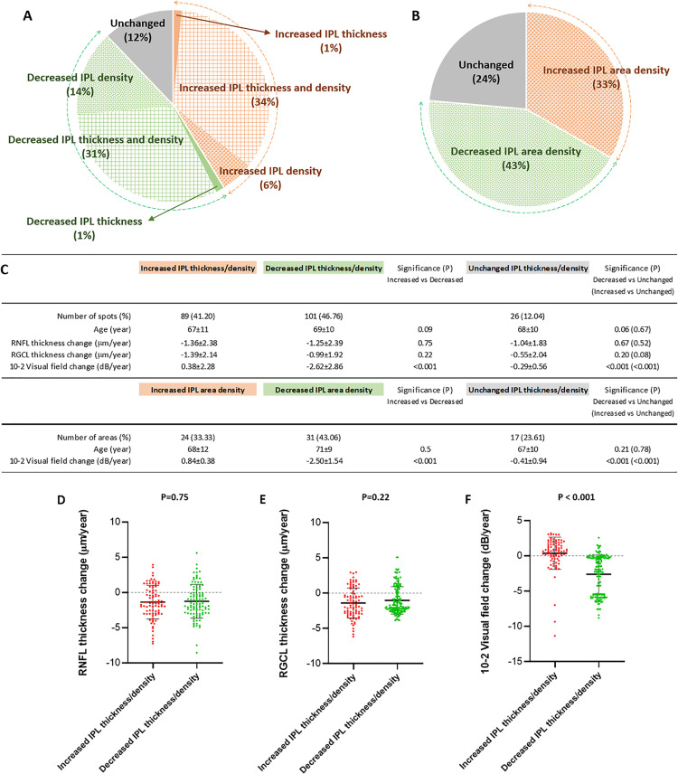
Glaucoma is a chronic neurodegenerative disease of the optic nerve and a leading cause of irreversible blindness, worldwide. While the experimental research using animal models provides growing information about cellular and molecular processes, parallel analysis of the clinical presentation of glaucoma accelerates the translational progress towards improved understanding, treatment, and clinical testing of glaucoma. Optic nerve axon injury triggers early alterations of retinal ganglion cell (RGC) synapses with function deficits prior to manifest RGC loss in animal models of glaucoma. For testing the clinical relevance of experimental observations, this study analyzed the functional correlation of localized alterations in the inner plexiform layer (IPL), where RGCs establish synaptic connections with retinal bipolar and amacrine cells. Participants of the study included a retrospective cohort of 36 eyes with glaucoma and a control group of 18 non-glaucomatous subjects followed for two-years. The IPL was analyzed on consecutively collected macular SD-OCT scans, and functional correlations with corresponding 10-2 visual field scores were tested using generalized estimating equations (GEE) models. The GEE-estimated rate of decrease in IPL thickness (R = 0.36, P<0.001) and IPL density (R = 0.36, P<0.001), as opposed to unchanged or increased IPL thickness or density, was significantly associated with visual field worsening at corresponding analysis locations. Based on multivariate logistic regression analysis, this association was independent from the patients' age, the baseline visual field scores, or the baseline thickness or alterations of retinal nerve fiber or RGC layers (P>0.05). These findings support early localized IPL alterations in correlation with progressing visual field defects in glaucomatous eyes. Considering the experimental data, glaucoma-related increase in IPL thickness/density might reflect dendritic remodeling, mitochondrial redistribution, and glial responses for synapse maintenance, but decreased IPL thickness/density might correspond to dendrite atrophy. The bridging of experimental data with clinical findings encourages further research along the translational path.
青光眼是一种慢性视神经退行性疾病,也是全球范围内导致不可逆转失明的主要原因。虽然使用动物模型的实验研究提供了越来越多的关于细胞和分子过程的信息,但对青光眼临床表现的平行分析加速了向更好地理解、治疗和临床测试青光眼的转化进展。视神经轴突损伤引发视网膜神经节细胞(RGC)突触的早期改变,在动物青光眼模型中表现出 RGC 丧失之前出现功能缺陷。为了测试实验观察的临床相关性,本研究分析了内丛状层(IPL)中局部改变的功能相关性,RGC 在此处与视网膜双极细胞和无长突细胞建立突触连接。该研究的参与者包括一个回顾性队列,其中包括 36 只青光眼眼和 18 只非青光眼对照组,随访时间为两年。在连续采集的黄斑 SD-OCT 扫描上分析 IPL,并使用广义估计方程(GEE)模型测试与相应的 10-2 视野分数的功能相关性。GEE 估计的 IPL 厚度(R = 0.36,P<0.001)和 IPL 密度(R = 0.36,P<0.001)的下降率,与相应分析位置的视野恶化显著相关。基于多元逻辑回归分析,这种相关性独立于患者的年龄、基线视野评分、或基线视网膜神经纤维或 RGC 层的厚度或改变(P>0.05)。这些发现支持在青光眼眼中与进行性视野缺陷相关的早期局部 IPL 改变。考虑到实验数据,与青光眼相关的 IPL 厚度/密度增加可能反映了树突重塑、线粒体再分布和胶质细胞对突触维持的反应,但 IPL 厚度/密度降低可能对应于树突萎缩。将实验数据与临床发现联系起来,鼓励沿着转化途径进一步研究。