Medical Physics and Biomedical Engineering Department, School of Medicine, Tehran University of Medical Sciences, 1417613151, Tehran, Iran.
Department of Stem Cells and Developmental Biology, Cell Science Research Center, Royan Institute for Stem Cell Biology and Technology, ACECR, Banihashem St., Resalat Highway, P.O. Box: 16635-148, 1665659911, Tehran, Iran.
BMC Med Imaging. 2021 Feb 25;21(1):37. doi: 10.1186/s12880-021-00562-y.
Intraoperative coronary angiography can tremendously reduce early coronary bypass graft failures. Fluorescent cardiac imaging provides an advanced method for intraoperative observation and real-time quantitation of blood flow with high resolution.
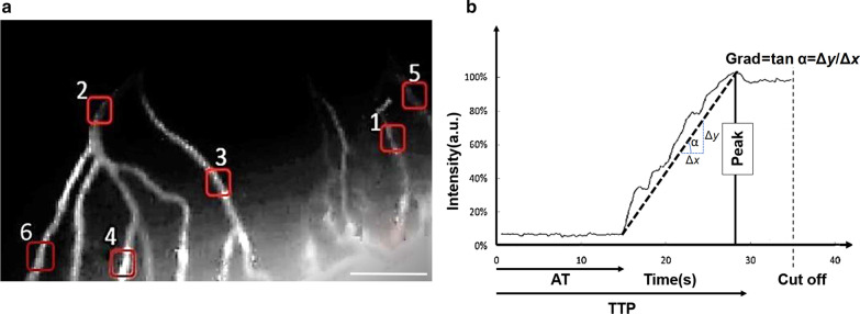
We devised a system comprised of an LED light source, special filters, lenses and a detector for preclinical coronary artery angiography. The optical setup was implemented by using two achromatic doublet lenses, two positive meniscus lenses, a band-pass filter, a pinhole and a CCD sensor. The setup was optimized by Zemax software. Optical design was further challenged to obtain more parallel light beams, less diffusion and higher resolutions to levels as small as arterioles. Ex vivo rat hearts were prepared and coronary arteries were retrogradely perfused by indocyanine green (ICG). Video angiography was employed to assess blood flow and plot time-dependent fluorescence intensity curve (TIC). Quantitation of blood flow was performed by calculating either the gradient of TIC or area under curve. The correlation between blood flow and each calculated parameters was assessed and used to evaluate the quality of flow.
High-resolution images of flow in coronary arteries were obtained as precise as 62 µm vessel diameter, by our custom-made ICG angiography system. The gradient of TIC was 3.4-6.3 s, while the area under curve indicated 712-1282 s values which ultimately gained correlation coefficients of 0.9938 and 0.9951 with relative blood flow, respectively.
The present ICG angiography system may facilitate evaluation of blood flow in animal studies of myocardial infarction and coronary artery grafts intraoperatively.
术中冠状动脉造影术可以极大地降低早期冠状动脉搭桥失败的风险。荧光心脏成像提供了一种先进的方法,用于术中观察和实时定量血流,具有高分辨率。
我们设计了一个由 LED 光源、特殊滤波器、透镜和探测器组成的系统,用于临床前冠状动脉血管造影。光学设置通过使用两个消色差双凸透镜、两个正弯月透镜、带通滤波器、针孔和 CCD 传感器来实现。通过 Zemax 软件对该系统进行了优化。光学设计进一步挑战在于获得更平行的光束、更少的扩散和更高的分辨率,达到甚至像小动脉这样的水平。通过吲哚菁绿(ICG)对离体大鼠心脏进行逆行灌注,制备离体大鼠心脏,并对冠状动脉进行逆行灌注。采用视频血管造影术评估血流,并绘制时间依赖性荧光强度曲线(TIC)。通过计算 TIC 的梯度或曲线下面积来进行血流定量。评估了血流与每个计算参数之间的相关性,并用于评估血流质量。
通过我们定制的 ICG 血管造影系统,获得了精确到 62µm 血管直径的冠状动脉内血流高分辨率图像。TIC 的梯度为 3.4-6.3s,而曲线下面积则分别指示 712-1282s 的值,最终与相对血流分别获得 0.9938 和 0.9951 的相关系数。
本研究中的 ICG 血管造影系统可有助于评估心肌梗死和冠状动脉旁路移植术中动物研究的血流情况。