Zötterman Johan, Tesselaar Erik, Elawa Sherif, Elmasry Moustafa, Farnebo Simon
From the Department of Hand and Plastic Surgery and Burns and Department of Clinical and Experimental Medicine, Linköping University, Linköping, Sweden.
Department of Medical Radiation Physics and Department of Clinical and Experimental Medicine, Linköping University, Linköping, Sweden.
Plast Reconstr Surg Glob Open. 2024 Jul 26;12(7):e5964. doi: 10.1097/GOX.0000000000005964. eCollection 2024 Jul.
Indocyanine green fluorescence angiography (ICG-FA) is often used for assessing tissue circulation in reconstructive surgery. Indocyanine green (ICG) is injected intravenously and visualized in the tissue with an infrared camera. The information is used to plan the surgery, for example, in free flap breast reconstructions. Laser speckle contrast imaging (LSCI) is another method that uses laser to assess tissue perfusion in the skin. Unlike ICG-FA, LSCI is noninvasive and may therefore have an advantaged compared with ICG-FA. The aim of this study was to evaluate the correlation between information obtained from these two techniques.
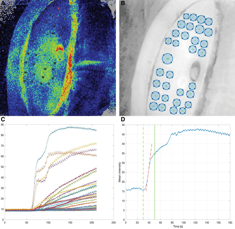
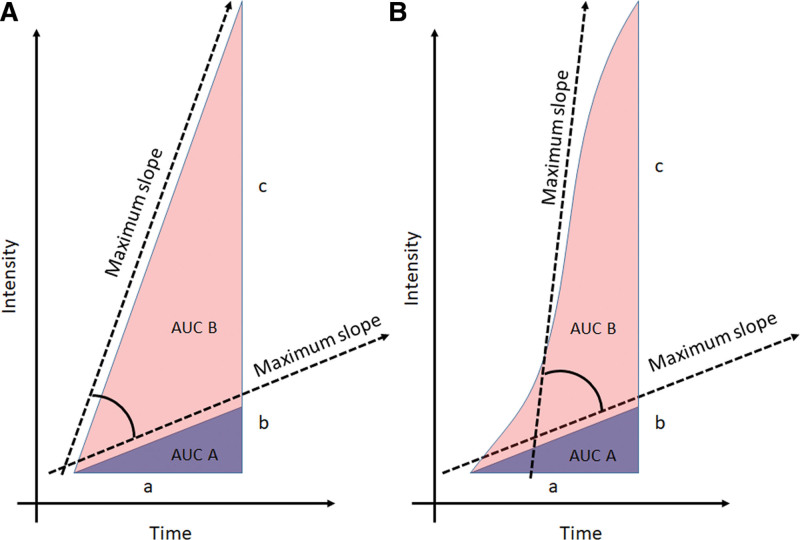
Five deep inferior epigastric perforator patients were included. The flaps were assessed with LSCI and ICG-FA. For LSCI, the perfusion was calculated in 32 regions of interest. For ICG-FA, the maximum slope and area under curve (AUC) were calculated based on average pixel intensity data.
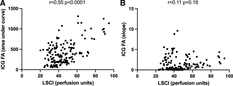
Large variations in maximum slope values could be seen between flaps, whereas AUC had lower variability within the same flap and between flaps. Pearson rank correlation comparing average perfusion (LSCI) and AUC (ICG-FA) showed a correlation between the values (r = 0.55, < 0.0001). No significant correlation was observed between perfusion and maximum slope (r = 0.11, = 0.18).
There is a significant correlation between data obtained using LSCI and ICG-FA, when ICG-FA data are presented as AUC of the ICG-FA intensity curve. Maximum slope lacks significant correlation with flap data obtained with LSCI. The study indicates that LSCI may be used in reconstructive surgery to assess tissue circulation in a way similar to ICG-FA.
吲哚菁绿荧光血管造影术(ICG-FA)常用于重建手术中评估组织循环。静脉注射吲哚菁绿(ICG),并用红外相机在组织中进行可视化观察。该信息用于手术规划,例如在游离皮瓣乳房重建术中。激光散斑对比成像(LSCI)是另一种利用激光评估皮肤组织灌注的方法。与ICG-FA不同,LSCI是非侵入性的,因此与ICG-FA相比可能具有优势。本研究的目的是评估从这两种技术获得的信息之间的相关性。
纳入5例腹壁下深动脉穿支皮瓣患者。使用LSCI和ICG-FA对皮瓣进行评估。对于LSCI,在32个感兴趣区域计算灌注情况。对于ICG-FA,基于平均像素强度数据计算最大斜率和曲线下面积(AUC)。
各皮瓣之间最大斜率值存在较大差异,而同一皮瓣内以及不同皮瓣之间AUC的变异性较低。比较平均灌注(LSCI)和AUC(ICG-FA)的Pearson等级相关性显示这些值之间存在相关性(r = 0.55,P < 0.0001)。未观察到灌注与最大斜率之间存在显著相关性(r = 0.11,P = 0.18)。
当将ICG-FA数据表示为ICG-FA强度曲线的AUC时,使用LSCI和ICG-FA获得的数据之间存在显著相关性。最大斜率与通过LSCI获得的皮瓣数据缺乏显著相关性。该研究表明,LSCI可用于重建手术中以类似于ICG-FA的方式评估组织循环。