Department of Clinical Medicine and Surgery, Section of Dermatology and Venereology, University of Naples Federico II, Via Pansini 5, Napoli, Italy.
Department of Neurosciences, Intensive Care Unit, Reproductive and Odontostomatological Sciences, University of Naples Federico II, Via Pansini 5, Napoli, Italy.
Diagn Pathol. 2021 Feb 25;16(1):16. doi: 10.1186/s13000-021-01075-6.
To date, very few studies on clinical-histopathological correlations of cutaneous disorders associated with COVID-19 have been conducted.
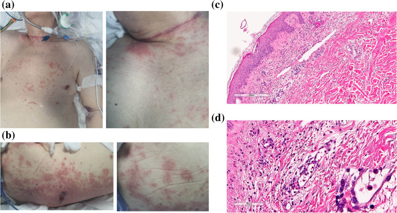
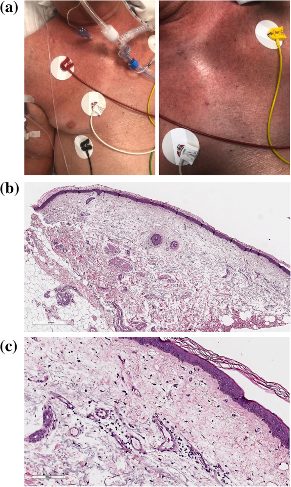
The Case 1 was a 90-year-old man, who tested positive for SARS-CoV-2 from a nasopharyngeal swab. Two days later, he was hospitalized and after eleven days transferred to Intensive Care Unit. A chest CT showed bilateral ground-glass opacities. Just that day, an erythematous maculo-papular rash appeared on trunk, shoulders and neck, becoming purpuric after few days. Histological evaluations revealed a chronic superficial dermatitis with purpuric aspects. The superficial and papillary dermis appeared edematous, with a perivascular lympho-granulocytic infiltrate and erythrocytic extravasation. At intraepithelial level, spongiosis and a granulocyte infiltrate were detected. Arterioles, capillaries and post-capillary venules showed endothelial swelling and appeared ectatic. The patient was treated with hydroxychloroquine, azithromycin, lopinavir-ritonavir and tocilizumab. Regrettably, due to severe lung impairment, he died. The Case 2 was a 85-year-old man, admitted to Intensive Care Unit, where he was intubated. He had tested positive for SARS-CoV-2 from a nasopharyngeal swab two days before. A chest RX showed bilateral atypical pneumonia. After seven days, a cutaneous reddening involving trunk, upper limbs, neck and face developed, configuring a sub-erythroderma. Histological evaluations displayed edema in the papillary and superficial reticular dermis, and a perivascular lymphocytic infiltrate in the superficial dermis. The patient was treated with hydroxychloroquine, azithromycin, lopinavir-ritonavir and tocilizumab. Sub-erythroderma as well as respiratory symptoms gradually improved until healing.
The endothelial swelling detected in the Case 1 could be a morphological expression of SARS-CoV-2-induced endothelial dysfunction. We hypothesize that cutaneous damage could be initiated by endothelial dysfunction, caused by SARS-CoV-2 infection of endothelial cells or induced by immune system activation. The disruption of endothelial integrity could enhance microvascular permeability, extravasation of inflammatory cells and cytokines, with cutaneous injury. The Case 2 developed a sub-erythroderma associated with COVID-19, and a non-specific chronic dermatitis was detected at histological level. We speculate that a purpuric rash could represent the cutaneous sign of a more severe coagulopathy, as highlighted histologically by vascular abnormalities, while a sub-erythroderma could be expression of viral hematogenous spreading, inducing a non-specific chronic dermatitis.
迄今为止,关于与 COVID-19 相关的皮肤疾病的临床-组织病理学相关性的研究很少。
病例 1 为一名 90 岁男性,从鼻咽拭子中检测到 SARS-CoV-2 呈阳性。两天后,他住院并在 11 天后转入重症监护病房。胸部 CT 显示双侧磨玻璃样混浊。就在那天,躯干、肩部和颈部出现了红斑性斑丘疹,几天后变成了紫癜。组织学评估显示为慢性浅表性皮炎伴紫癜表现。浅层和乳头状真皮出现水肿,伴有血管周围淋巴-粒细胞浸润和红细胞外渗。在上皮内水平,可见海绵状变性和粒细胞浸润。小动脉、毛细血管和小静脉后小静脉显示内皮肿胀并呈扩张状态。该患者接受了羟氯喹、阿奇霉素、洛匹那韦-利托那韦和托珠单抗治疗。遗憾的是,由于严重的肺部损伤,他去世了。病例 2 为一名 85 岁男性,因 SARS-CoV-2 检测呈阳性,被收入重症监护病房并插管。他在两天前从鼻咽拭子中检测到 SARS-CoV-2 呈阳性。胸部 RX 显示双侧非典型肺炎。七天后,躯干、上肢、颈部和面部出现皮肤变红,形成亚红皮病。组织学评估显示真皮乳头和浅层网状真皮水肿,浅层真皮中血管周围淋巴细胞浸润。该患者接受了羟氯喹、阿奇霉素、洛匹那韦-利托那韦和托珠单抗治疗。亚红皮病和呼吸道症状逐渐改善直至痊愈。
病例 1 中检测到的内皮肿胀可能是 SARS-CoV-2 诱导的内皮功能障碍的形态学表现。我们假设,皮肤损伤可能是由 SARS-CoV-2 感染内皮细胞或由免疫系统激活引起的内皮功能障碍引发的。内皮完整性的破坏可增强微血管通透性、炎症细胞和细胞因子外渗,导致皮肤损伤。病例 2 出现与 COVID-19 相关的亚红皮病,组织学水平发现非特异性慢性皮炎。我们推测,瘀斑可能代表更严重的凝血功能障碍的皮肤征象,如血管异常在组织学上突出显示的那样,而亚红皮病可能是病毒血行播散的表现,引起非特异性慢性皮炎。