POLARIS, Imaging Sciences, Department of Infection, Immunity and Cardiovascular Disease, University of Sheffield, Sheffield, United Kingdom.
POLARIS, Imaging Sciences, Department of Infection, Immunity and Cardiovascular Disease, University of Sheffield, Sheffield, United Kingdom.
Prog Nucl Magn Reson Spectrosc. 2021 Feb;122:42-62. doi: 10.1016/j.pnmrs.2020.11.002. Epub 2020 Dec 9.
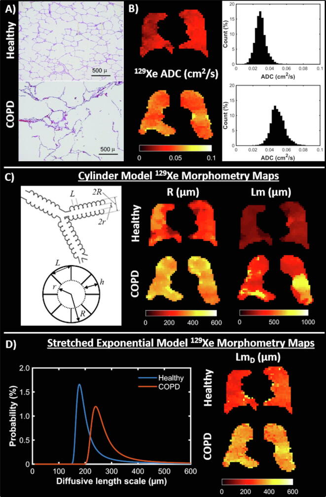
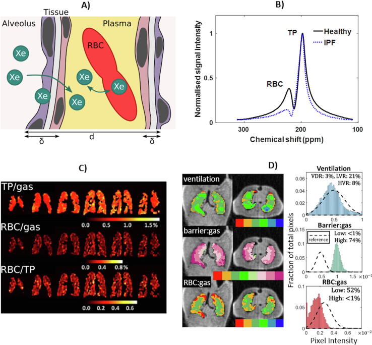
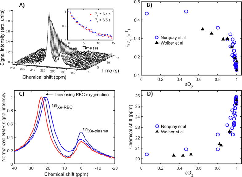
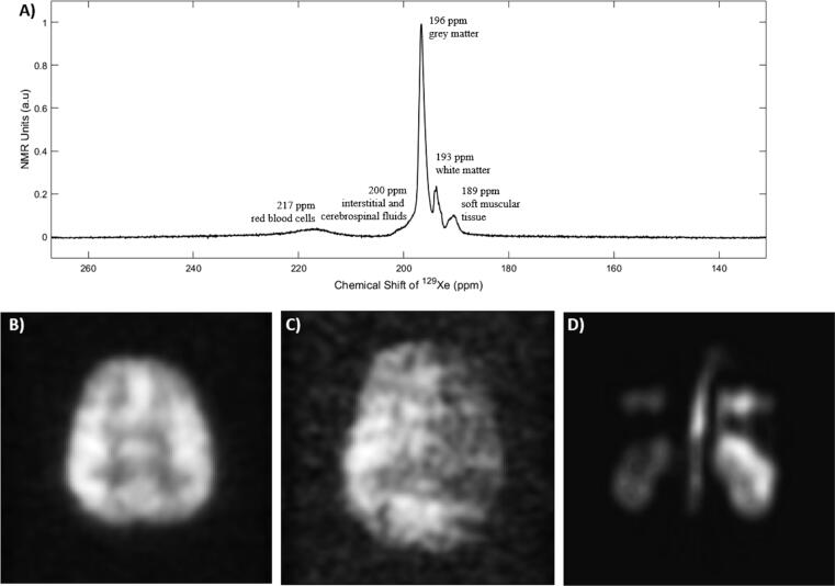
Hyperpolarised gas lung MRI using xenon-129 can provide detailed 3D images of the ventilated lung airspaces, and can be applied to quantify lung microstructure and detailed aspects of lung function such as gas exchange. It is sensitive to functional and structural changes in early lung disease and can be used in longitudinal studies of disease progression and therapy response. The ability of Xe to dissolve into the blood stream and its chemical shift sensitivity to its local environment allow monitoring of gas exchange in the lungs, perfusion of the brain and kidneys, and blood oxygenation. This article reviews the methods and applications of in vivoXe MR in humans, with a focus on the physics of polarisation by optical pumping, radiofrequency coil and pulse sequence design, and the in vivo applications of Xe MRI and MRS to examine lung ventilation, microstructure and gas exchange, blood oxygenation, and perfusion of the brain and kidneys.
利用氙气-129 进行超极化气体肺部 MRI 可以提供通气肺部气腔的详细 3D 图像,并可用于量化肺部微观结构和肺部功能的详细方面,例如气体交换。它对早期肺部疾病的功能和结构变化敏感,可用于疾病进展和治疗反应的纵向研究。氙气溶解到血流中的能力及其对局部环境的化学位移敏感性允许监测肺部的气体交换、大脑和肾脏的灌注以及血氧。本文综述了活体 Xe MR 在人体中的方法和应用,重点介绍了光泵浦、射频线圈和脉冲序列设计的极化物理,以及 Xe MRI 和 MRS 在检查肺部通气、微观结构和气体交换、血氧和大脑及肾脏灌注方面的活体应用。