Department of Translational Molecular Pathology, The University of Texas MD Anderson Cancer Center, Unit 9512130 Holcombe Blvd, Houston, TX, 77030, USA.
Departments of Thoracic and Cardiovascular Surgery, The University of Texas MD Anderson Cancer Center, Houston, TX, USA.
Sci Rep. 2021 Feb 25;11(1):4530. doi: 10.1038/s41598-021-83858-x.
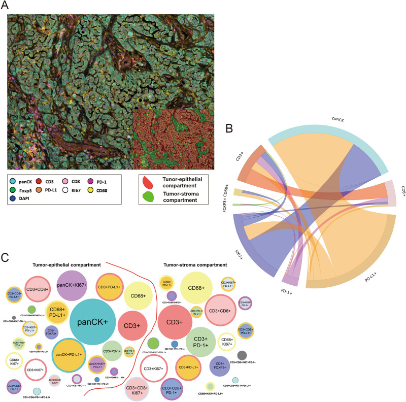
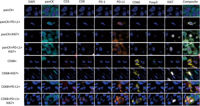
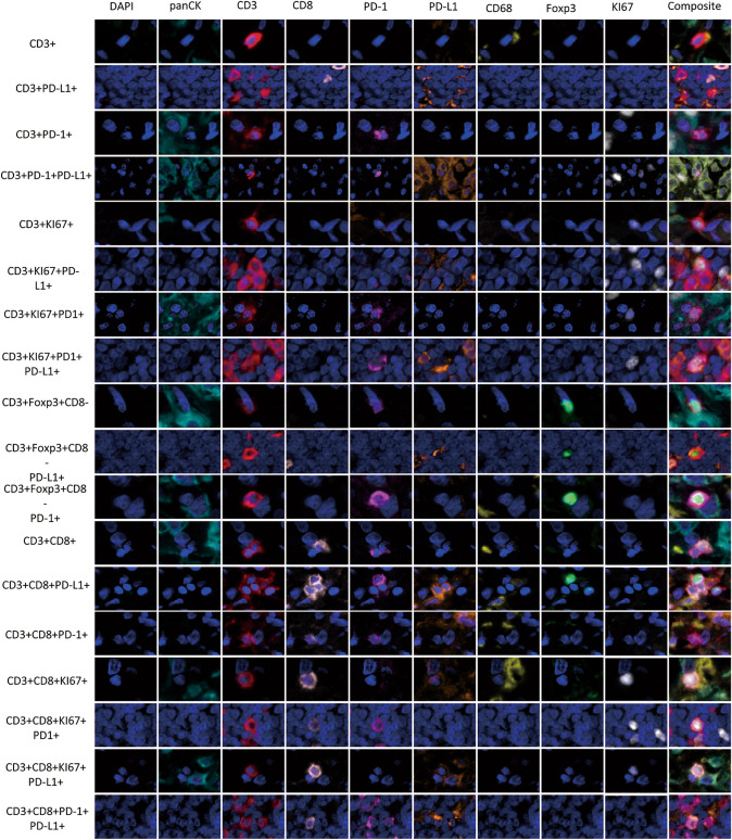
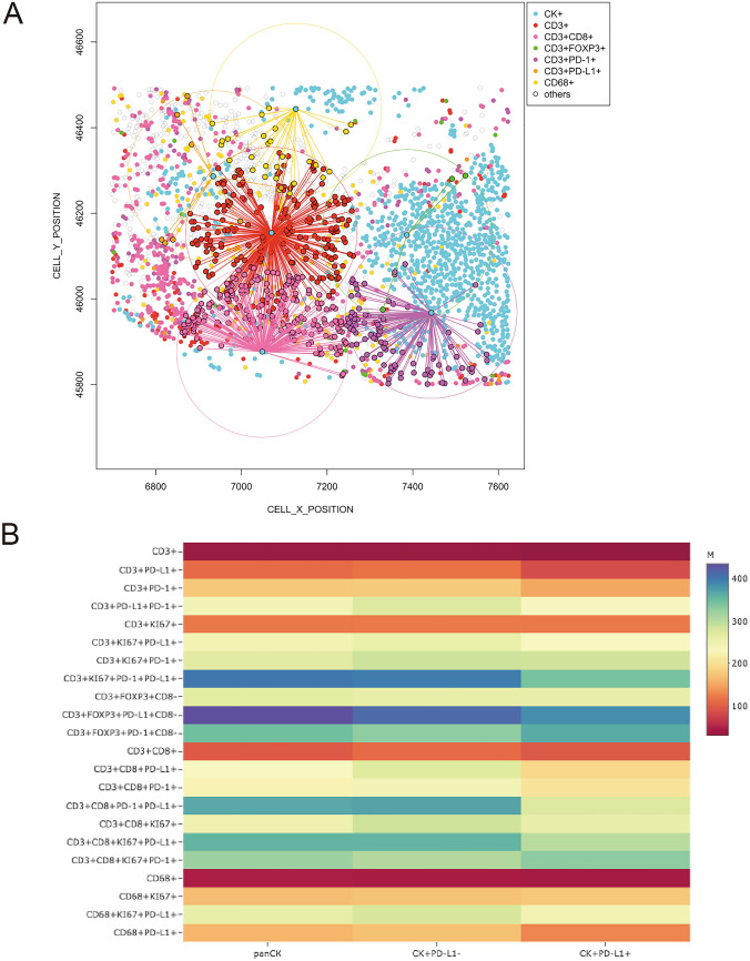
Immune profiling is becoming a vital tool for identifying predictive and prognostic markers for translational studies. The study of the tumor microenvironment (TME) in paraffin tumor tissues such as malignant pleural mesothelioma (MPM) could yield insights to actionable targets to improve patient outcome. Here, we optimized and tested a new immune-profiling method to characterize immune cell phenotypes in paraffin tissues and explore the co-localization and spatial distribution between the immune cells within the TME and the stromal or tumor compartments. Tonsil tissues and tissue microarray (TMA) were used to optimize an automated nine-color multiplex immunofluorescence (mIF) panel to study the TME using eight antibodies: PD-L1, PD-1, CD3, CD8, Foxp3, CD68, KI67, and pancytokeratin. To explore the potential role of the cells into the TME with this mIF panel we applied this panel in twelve MPM cases to assess the multiple cell phenotypes obtained from the image analysis and well as their spatial distribution in this cohort. We successful optimized and applied an automated nine-color mIF panel to explore a small set of MPM cases. Image analysis showed a high degree of cell phenotype diversity with immunosuppression patterns in the TME of the MPM cases. Mapping the geographic cell phenotype distribution in the TME, we were able to identify two distinct, complex immune landscapes characterized by specific patterns of cellular distribution as well as cell phenotype interactions with malignant cells. Successful we showed the optimization and reproducibility of our mIF panel and their incorporation for comprehensive TME immune profiling into translational studies that could refine our ability to correlate immunologic phenotypes with specific patterns of cells distribution and distance analysis. Overall, this will improve our ability to understand the behavior of cells within the TME and predict new treatment strategies to improve patient outcome.
免疫分析正成为识别转化研究中预测性和预后性标志物的重要工具。对石蜡肿瘤组织(如恶性胸膜间皮瘤)中的肿瘤微环境(TME)进行研究,可以深入了解可行的靶点,以改善患者的预后。在这里,我们优化并测试了一种新的免疫分析方法,以分析石蜡组织中的免疫细胞表型,并探索 TME 内免疫细胞与基质或肿瘤区室之间的共定位和空间分布。我们使用扁桃体组织和组织微阵列(TMA)优化了一种自动化的九色多重免疫荧光(mIF)检测 panel,使用八种抗体来研究 TME:PD-L1、PD-1、CD3、CD8、Foxp3、CD68、Ki67 和广谱细胞角蛋白。为了用该 mIF 检测 panel 探索细胞进入 TME 的潜在作用,我们在 12 例 MPM 病例中应用了该 panel,以评估从图像分析中获得的多种细胞表型,以及它们在该队列中的空间分布。我们成功优化并应用了自动化的九色 mIF 检测 panel 来探索一小部分 MPM 病例。图像分析显示,TME 中的 MPM 病例具有高度的细胞表型多样性和免疫抑制模式。通过对 TME 中细胞表型分布的地理定位,我们能够识别出两种不同的、复杂的免疫景观,其特征是特定的细胞分布模式以及细胞表型与恶性细胞的相互作用。我们成功地展示了我们的 mIF 检测 panel 的优化和重现性,以及它们在转化研究中对全面的 TME 免疫分析的纳入,这可以提高我们将免疫表型与特定的细胞分布模式和距离分析相关联的能力。总的来说,这将提高我们理解 TME 中细胞行为的能力,并预测新的治疗策略,以改善患者的预后。