Narayanan Priya Lakshmi, Raza Shan E Ahmed, Hall Allison H, Marks Jeffrey R, King Lorraine, West Robert B, Hernandez Lucia, Guppy Naomi, Dowsett Mitch, Gusterson Barry, Maley Carlo, Hwang E Shelley, Yuan Yinyin
Centre for Evolution and Cancer, Institute of Cancer Research, London, UK.
Division of Molecular Pathology, Institute of Cancer Research, London, UK.
NPJ Breast Cancer. 2021 Mar 1;7(1):19. doi: 10.1038/s41523-020-00205-5.
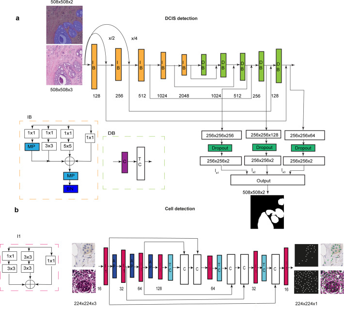
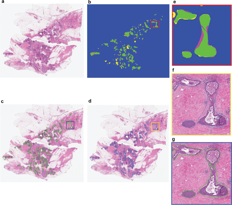
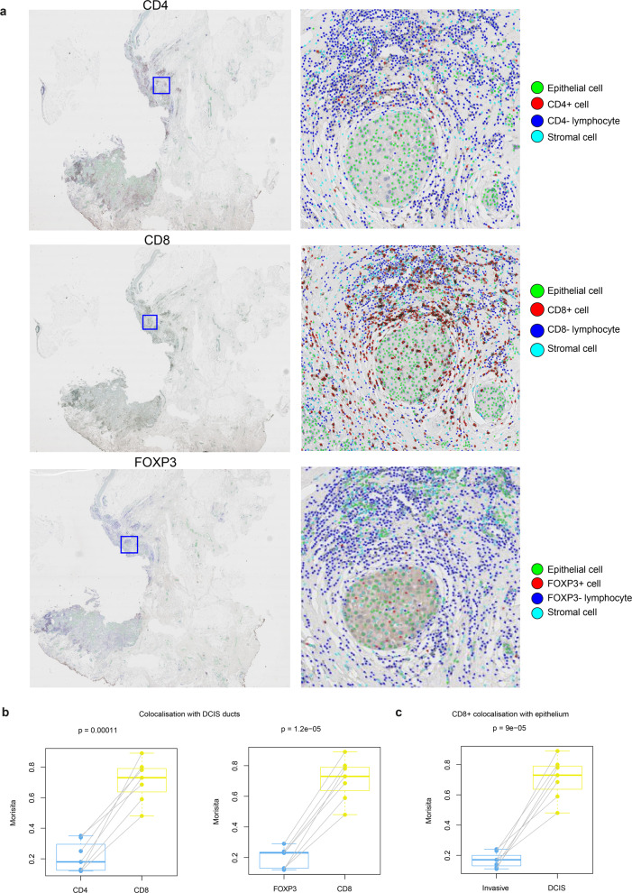
Despite increasing evidence supporting the clinical relevance of tumour infiltrating lymphocytes (TILs) in invasive breast cancer, TIL spatial variability within ductal carcinoma in situ (DCIS) samples and its association with progression are not well understood. To characterise tissue spatial architecture and the microenvironment of DCIS, we designed and validated a new deep learning pipeline, UNMaSk. Following automated detection of individual DCIS ducts using a new method IM-Net, we applied spatial tessellation to create virtual boundaries for each duct. To study local TIL infiltration for each duct, DRDIN was developed for mapping the distribution of TILs. In a dataset comprising grade 2-3 pure DCIS and DCIS adjacent to invasive cancer (adjacent DCIS), we found that pure DCIS cases had more TILs compared to adjacent DCIS. However, the colocalisation of TILs with DCIS ducts was significantly lower in pure DCIS compared to adjacent DCIS, which may suggest a more inflamed tissue ecology local to DCIS ducts in adjacent DCIS cases. Our study demonstrates that technological developments in deep convolutional neural networks and digital pathology can enable an automated morphological and microenvironmental analysis of DCIS, providing a new way to study differential immune ecology for individual ducts and identify new markers of progression.
尽管越来越多的证据支持肿瘤浸润淋巴细胞(TILs)在浸润性乳腺癌中的临床相关性,但导管原位癌(DCIS)样本中TIL的空间变异性及其与进展的关联仍未得到充分了解。为了表征DCIS的组织空间结构和微环境,我们设计并验证了一种新的深度学习管道UNMaSk。在使用新方法IM-Net自动检测单个DCIS导管后,我们应用空间镶嵌为每个导管创建虚拟边界。为了研究每个导管的局部TIL浸润情况,我们开发了DRDIN来绘制TIL的分布。在一个包含2-3级纯DCIS和浸润性癌旁DCIS(相邻DCIS)的数据集里,我们发现纯DCIS病例的TIL比相邻DCIS更多。然而,与相邻DCIS相比,纯DCIS中TIL与DCIS导管的共定位显著更低,这可能表明相邻DCIS病例中DCIS导管局部的组织生态炎症更严重。我们的研究表明,深度卷积神经网络和数字病理学的技术发展能够对DCIS进行自动形态学和微环境分析,可以为研究单个导管的差异免疫生态和识别新的进展标志物提供新方法。