Functional Neuroimaging Unit, University of Montreal Geriatric Institute, 4565, Queen-Mary Road, Montreal, QC, H3W 1W5, Canada.
Center for Advanced Research in Sleep Medicine (CARSM), Hôpital du Sacré-Cœur de Montréal, 5400 Gouin Boulevard West, Montreal, QC, H4J 1C5, Canada.
Sci Rep. 2021 Mar 1;11(1):4905. doi: 10.1038/s41598-021-84417-0.
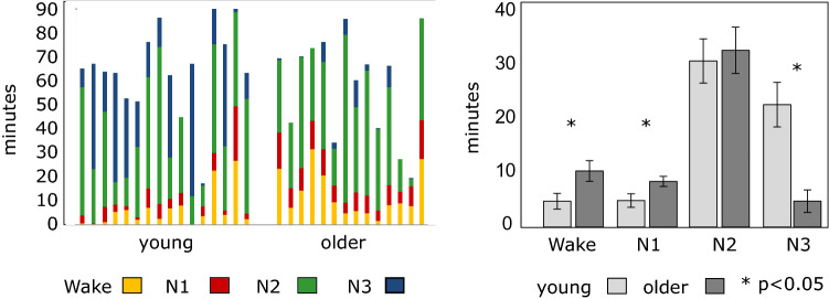
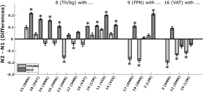
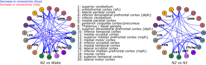
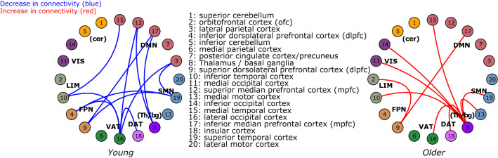
Even though sleep modification is a hallmark of the aging process, age-related changes in functional connectivity using functional Magnetic Resonance Imaging (fMRI) during sleep, remain unknown. Here, we combined electroencephalography and fMRI to examine functional connectivity differences between wakefulness and light sleep stages (N1 and N2 stages) in 16 young (23.1 ± 3.3y; 7 women), and 14 older individuals (59.6 ± 5.7y; 8 women). Results revealed extended, distributed (inter-between) and local (intra-within) decreases in network connectivity during sleep both in young and older individuals. However, compared to the young participants, older individuals showed lower decreases in connectivity or even increases in connectivity between thalamus/basal ganglia and several cerebral regions as well as between frontal regions of various networks. These findings reflect a reduced ability of the older brain to disconnect during sleep that may impede optimal disengagement for loss of responsiveness, enhanced lighter and fragmented sleep, and contribute to age effects on sleep-dependent brain plasticity.
尽管睡眠的改变是衰老过程的一个标志,但在睡眠过程中使用功能磁共振成像 (fMRI) 研究与年龄相关的功能连接变化仍然未知。在这里,我们结合脑电图和 fMRI 检查了 16 名年轻(23.1 ± 3.3 岁;7 名女性)和 14 名老年人(59.6 ± 5.7 岁;8 名女性)在清醒和轻度睡眠阶段(N1 和 N2 阶段)之间的功能连接差异。结果显示,在年轻人和老年人中,睡眠期间网络连接的扩展、分布式(间内)和局部(内)都出现了减少。然而,与年轻参与者相比,老年人在丘脑/基底神经节与几个大脑区域之间以及各个网络的额叶区域之间的连接减少较低,甚至出现连接增加。这些发现反映了老年人大脑在睡眠期间断开连接的能力降低,这可能会阻碍对反应能力丧失的最佳脱离,增强更轻和碎片化的睡眠,并导致年龄对睡眠相关脑可塑性的影响。