Department of Ophthalmology, Visual and Anatomical Sciences, Wayne State University School of Medicine, Detroit, Michigan, United States of America.
Beaumont Research Institute, Beaumont Health, Royal Oak, Michigan, United States of America.
PLoS One. 2021 Mar 4;16(3):e0245161. doi: 10.1371/journal.pone.0245161. eCollection 2021.
The phosphodiesterase inhibitor sildenafil is a promising treatment for neurodegenerative disease, but it can cause oxidative stress in photoreceptors ex vivo and degrade visual performance in humans. Here, we test the hypotheses that in wildtype mice sildenafil causes i) wide-spread photoreceptor oxidative stress in vivo that is linked with ii) impaired vision.
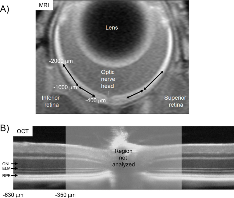
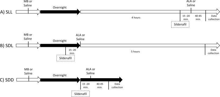
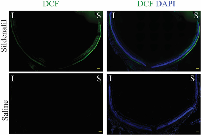
In dark or light-adapted C57BL/6 mice ± sildenafil treatment, the presence of oxidative stress was evaluated in retina laminae in vivo by QUEnch-assiSTed (QUEST) magnetic resonance imaging, in the subretinal space in vivo by QUEST optical coherence tomography, and in freshly excised retina by a dichlorofluorescein assay. Visual performance indices were also evaluated by QUEST optokinetic tracking.
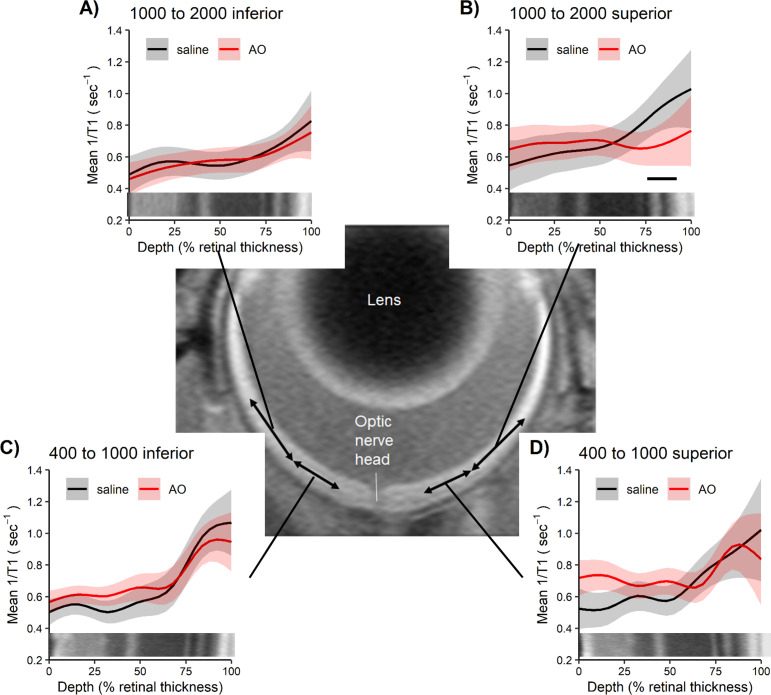
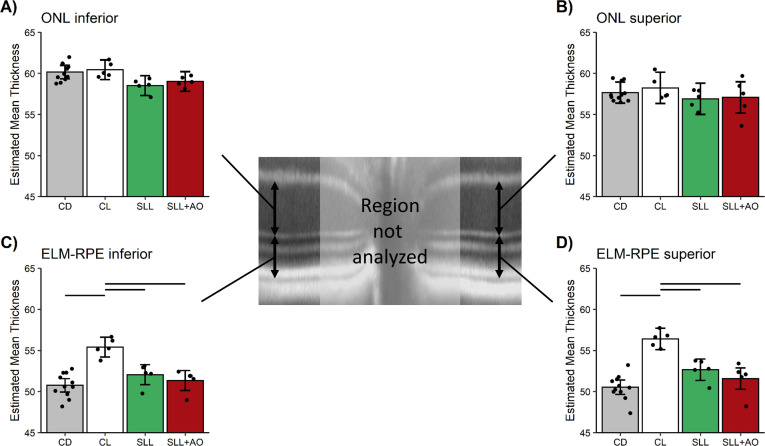
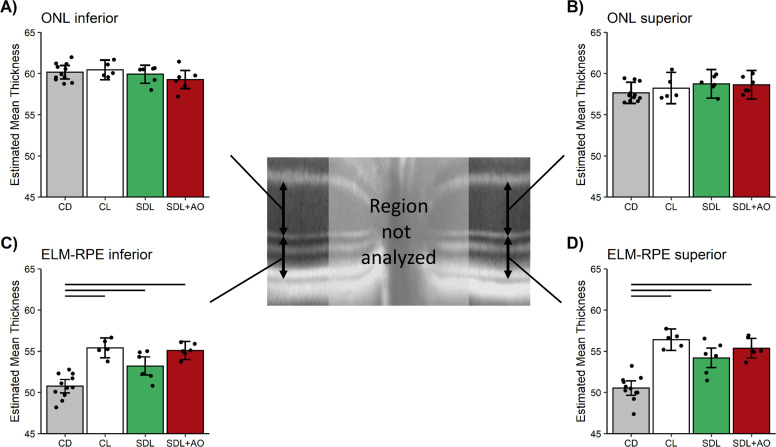
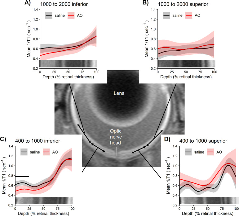
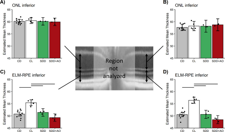
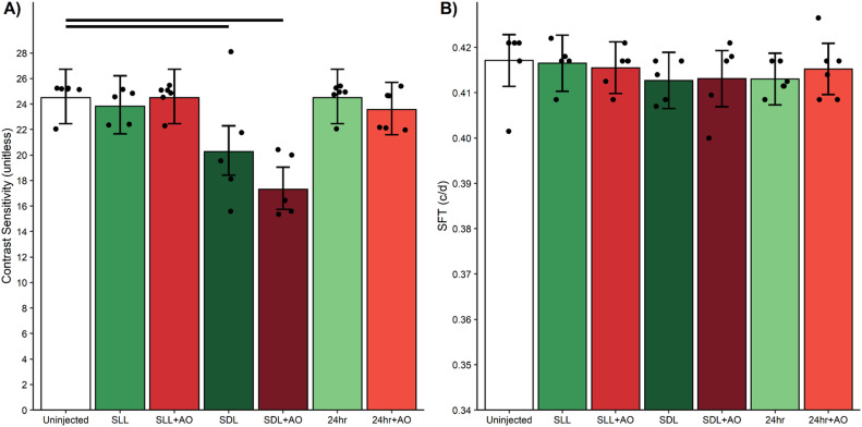
In light-adapted mice, 1 hr post-sildenafil administration, oxidative stress was most evident in the superior peripheral outer retina on both in vivo and ex vivo examinations; little evidence was noted for central retina oxidative stress in vivo and ex vivo. In dark-adapted mice 1 hr after sildenafil, no evidence for outer retina oxidative stress was found in vivo. Evidence for sildenafil-induced central retina rod cGMP accumulation was suggested as a panretinally thinner, dark-like subretinal space thickness in light-adapted mice at 1 hr but not 5 hr post-sildenafil. Cone-based visual performance was impaired by 5 hr post-sildenafil and not corrected with anti-oxidants; vision was normal at 1 hr and 24 hr post-sildenafil.
The sildenafil-induced spatiotemporal pattern of oxidative stress in photoreceptors dominated by rods was unrelated to impairment of cone-based visual performance in wildtype mice.
磷酸二酯酶抑制剂西地那非是治疗神经退行性疾病的一种有前途的方法,但它会在体外引起光感受器的氧化应激,并降低人类的视觉表现。在这里,我们测试了以下假设:在野生型小鼠中,西地那非会引起 i)广泛的光感受器体内氧化应激,这与 ii)视力受损有关。
在黑暗或光照适应的 C57BL/6 小鼠中,用 QUEnch-assiSTed(QUEST)磁共振成像评估视网膜层中的体内氧化应激,用 QUEST 光学相干断层扫描评估亚视网膜空间中的体内氧化应激,并用二氯荧光素测定法评估新鲜分离的视网膜。视觉表现指数也通过 QUEST 视动追踪进行评估。
在光照适应的小鼠中,西地那非给药后 1 小时,在体内和体外检查中,在外周上象限的外视网膜中观察到最明显的氧化应激;在体内和体外检查中,中央视网膜的氧化应激证据很少。在光照适应的黑暗适应小鼠中,在西地那非后 1 小时,在体内未发现外视网膜氧化应激的证据。在光照适应的小鼠中,在西地那非后 1 小时但不在 5 小时时,暗示存在由西地那非诱导的中心视网膜杆 cGMP 积累,表现为更薄的、类似暗的亚视网膜空间厚度。在西地那非后 5 小时,视锥细胞的视觉表现受损,而抗氧化剂无法纠正;在西地那非后 1 小时和 24 小时,视力正常。
在野生型小鼠中,光感受器中由杆状细胞主导的西地那非诱导的氧化应激时空模式与视锥细胞视觉表现受损无关。