Department of Ophthalmology, Visual and Anatomical Sciences, Wayne State University School of Medicine, 540 E. Canfield, Detroit, MI, 48201, USA.
Beaumont Research Institute, Beaumont Health, Royal Oak, MI, 48073, USA.
Geroscience. 2020 Apr;42(2):563-574. doi: 10.1007/s11357-020-00162-8. Epub 2020 Jan 25.
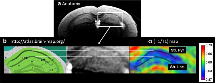
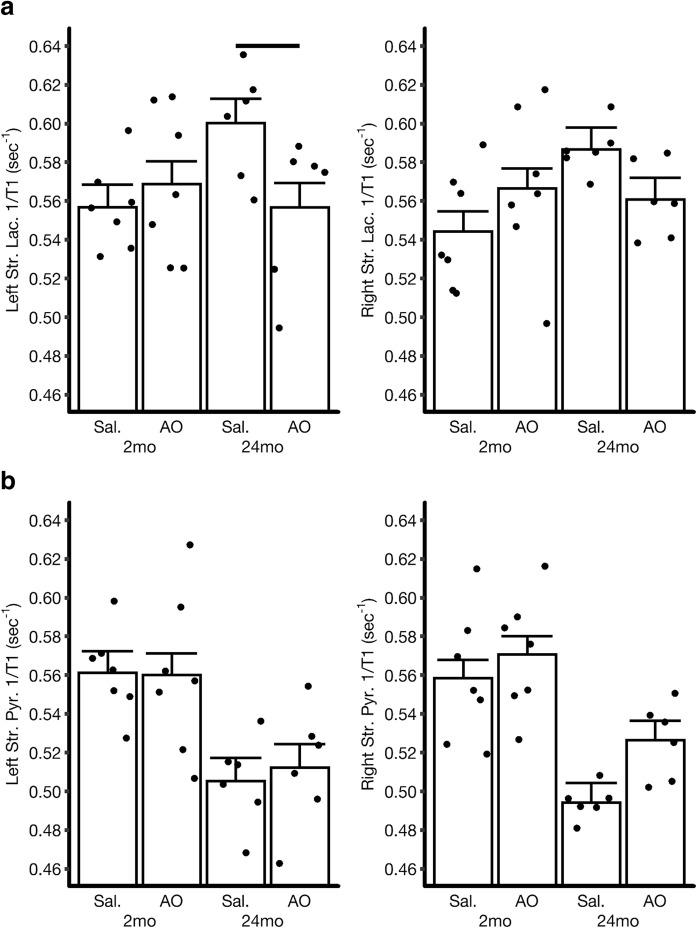
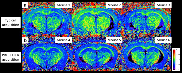
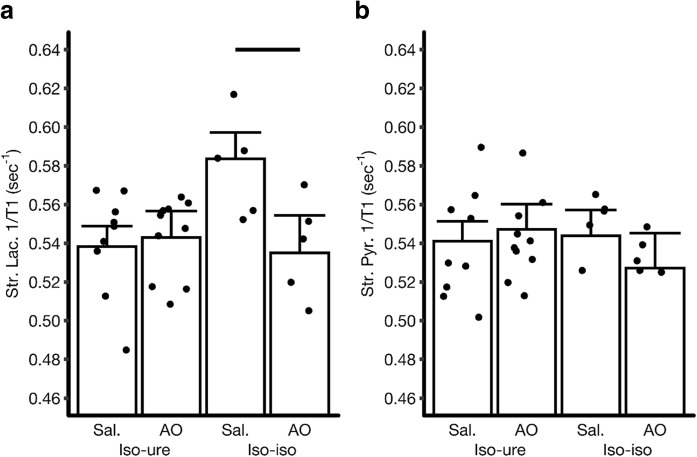
Age-related impairments in spatial learning and memory often precede non-familial neurodegenerative disease. Ex vivo studies suggest that physiologic age-related oxidative stress in hippocampus area CA1 may contribute to prodromal spatial disorientation and to morbidity. Yet, conventional blood or cerebrospinal fluid assays appear insufficient for early detection or management of oxidative stress within CA1 sub-regions in vivo. Here, we address this biomarker problem using a non-invasive MRI index of CA1 laminae oxidative stress based on reduction in R1 (= 1/T1) after anti-oxidant administration. An R1 reduction reflects quenching of continuous and excessive production of endogenous paramagnetic free radicals. Careful motion-correction image acquisition, and avoiding repeated exposure to isoflurane, facilitates detection of hippocampus CA1 laminae oxidative stress with QUEnch-assiSTed (QUEST) MRI. Intriguingly, age- and isoflurane-related oxidative stress is localized to the stratum lacunosum of the CA1 region. Our data raise the possibility of using QUEST MRI and FDA-approved anti-oxidants to remediate spatial disorientation and later neurodegeneration with age in animals and humans.
年龄相关性的空间学习和记忆损伤通常先于非家族性神经退行性疾病。离体研究表明,海马 CA1 区与生理年龄相关的氧化应激可能导致前驱性空间定向障碍和发病。然而,常规的血液或脑脊液检测似乎不足以在体内早期检测或管理 CA1 亚区的氧化应激。在这里,我们使用基于抗氧化剂给药后 R1(= 1/T1)减少的 CA1 层氧化应激的非侵入性 MRI 指数来解决这个生物标志物问题。R1 减少反映了连续和过度产生的内源性顺磁自由基的淬灭。仔细的运动校正图像采集,并避免反复暴露于异氟烷中,有助于使用 QUEnch-assiSTed(QUEST)MRI 检测海马 CA1 层的氧化应激。有趣的是,与年龄和异氟烷相关的氧化应激局限于 CA1 区域的腔隙层。我们的数据提出了使用 QUEST MRI 和 FDA 批准的抗氧化剂来改善动物和人类的空间定向障碍和随后的年龄相关性神经退行性变的可能性。