Department of Cardiology, Center for Translational Medicine, Institute of Precision Medicine, The First Affiliated Hospital, Sun Yat-sen University, Guangzhou 510080, China; NHC Key Laboratory of Assisted Circulation, Sun Yat-sen University, Guangzhou 510080, China.
Key Laboratory of Gene Engineering of the Ministry of Education, State Key Laboratory for Biocontrol, Sun Yat-sen University, Guangzhou 510275, China.
Mol Ther. 2021 Jul 7;29(7):2253-2267. doi: 10.1016/j.ymthe.2021.03.004. Epub 2021 Mar 5.
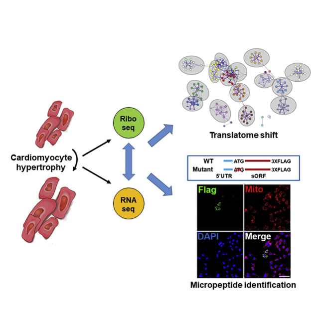
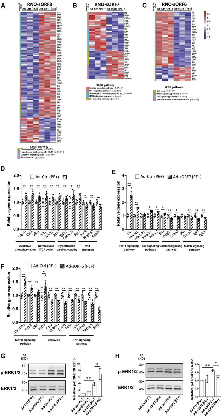
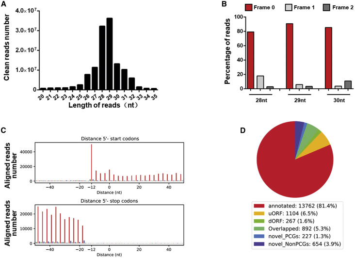
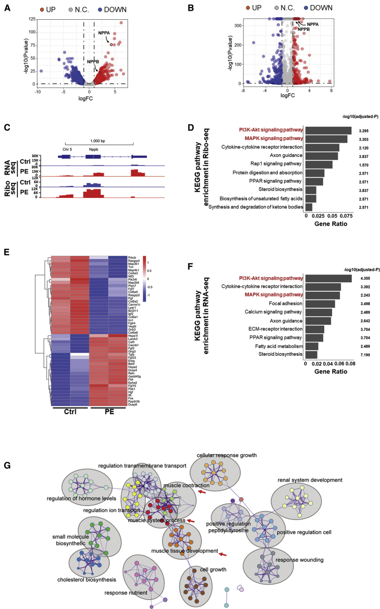
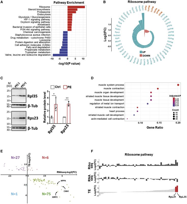
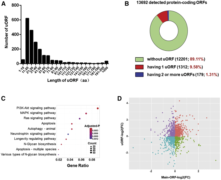
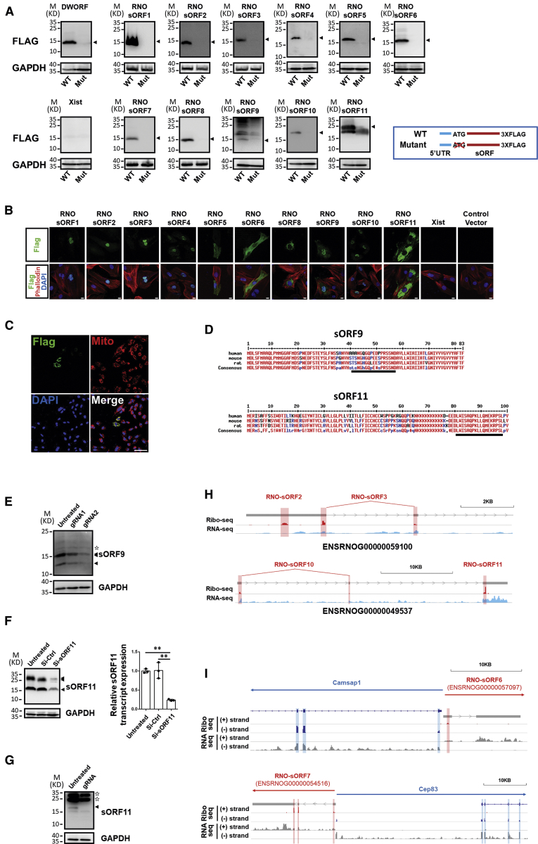
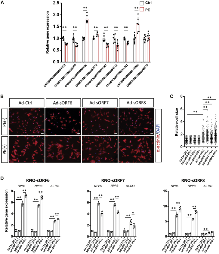
Hypertrophic growth of cardiomyocytes is one of the major compensatory responses in the heart after physiological or pathological stimulation. Protein synthesis enhancement, which is mediated by the translation of messenger RNAs, is one of the main features of cardiomyocyte hypertrophy. Although the transcriptome shift caused by cardiac hypertrophy induced by different stimuli has been extensively investigated, translatome dynamics in this cellular process has been less studied. Here, we generated a nucleotide-resolution translatome as well as transcriptome data from isolated primary cardiomyocytes undergoing hypertrophy. More than 10,000 open reading frames (ORFs) were detected from the deep sequencing of ribosome-protected fragments (Ribo-seq), which orchestrated the shift of the translatome in hypertrophied cardiomyocytes. Our data suggest that rather than increase the translational rate of ribosomes, the increased efficiency of protein synthesis in cardiomyocyte hypertrophy was attributable to an increased quantity of ribosomes. In addition, more than 100 uncharacterized short ORFs (sORFs) were detected in long noncoding RNA genes from Ribo-seq with potential of micropeptide coding. In a random test of 15 candidates, the coding potential of 11 sORFs was experimentally supported. Three micropeptides were identified to regulate cardiomyocyte hypertrophy by modulating the activities of oxidative phosphorylation, the calcium signaling pathway, and the mitogen-activated protein kinase (MAPK) pathway. Our study provides a genome-wide overview of the translational controls behind cardiomyocyte hypertrophy and demonstrates an unrecognized role of micropeptides in cardiomyocyte biology.
心肌细胞的肥厚生长是心脏在生理或病理刺激后主要的代偿反应之一。蛋白质合成的增强是心肌肥厚的主要特征之一,它是通过信使 RNA 的翻译来介导的。虽然不同刺激引起的心脏肥厚所导致的转录组变化已经得到了广泛的研究,但这个细胞过程中的翻译组动态研究较少。在这里,我们从经历肥厚的分离原代心肌细胞中生成了核苷酸分辨率的翻译组和转录组数据。核糖体保护片段(Ribo-seq)的深度测序检测到了超过 10000 个开放阅读框(ORFs),这些 ORFs 协调了肥厚心肌细胞中转录组的变化。我们的数据表明,心肌肥厚中蛋白质合成效率的提高不是通过增加核糖体的翻译速率,而是归因于核糖体数量的增加。此外,从 Ribo-seq 中的长非编码 RNA 基因中检测到超过 100 个未被表征的短开放阅读框(sORFs),这些 sORFs 具有微肽编码的潜力。在对 15 个候选者的随机测试中,有 11 个 sORFs 的编码潜力得到了实验支持。三个微肽被鉴定为通过调节氧化磷酸化、钙信号通路和丝裂原激活蛋白激酶(MAPK)通路来调节心肌肥厚。我们的研究提供了心肌肥厚背后翻译控制的全基因组概述,并证明了微肽在心肌生物学中的未被认识的作用。