Bi Suyan, Yuan Qingqing, Dai Zhitao, Sun Xingru, Wan Sohaimi Wan Fatihah Binti, Bin Yusoff Ahmad Lutfi
School of Medical Sciences, Universiti Sains Malaysia, Kelantan, Malaysia.
National Cancer Center/National Clinical Research Center for Cancer/ Cancer Hospital & Shenzhen Hospital, Chinese Academy of Medical Sciences and Peking Union Medical College, Shenzhen, China.
Front Oncol. 2024 Sep 2;14:1414337. doi: 10.3389/fonc.2024.1414337. eCollection 2024.
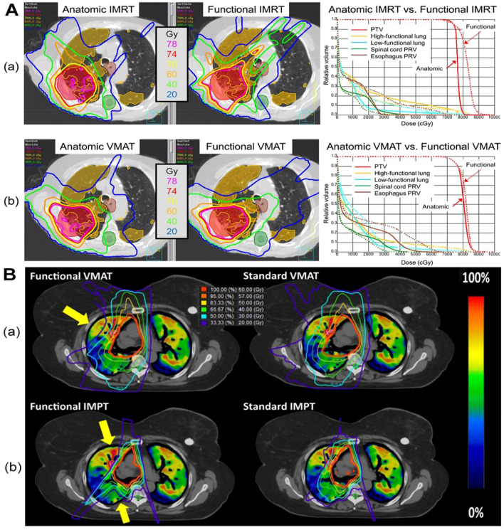
The objective of this review is to examine the potential benefits and challenges of CT-based lung function imaging in radiotherapy over recent decades. This includes reviewing background information, defining related concepts, classifying and reviewing existing studies, and proposing directions for further investigation. The lung function imaging techniques reviewed herein encompass CT-based methods, specifically utilizing phase-resolved four-dimensional CT (4D-CT) or end-inspiratory and end-expiratory CT scans, to delineate distinct functional regions within the lungs. These methods extract crucial functional parameters, including lung volume and ventilation distribution, pivotal for assessing and characterizing the functional capacity of the lungs. CT-based lung ventilation imaging offers numerous advantages, notably in the realm of thoracic radiotherapy. By utilizing routine CT scans, additional radiation exposure and financial burdens on patients can be avoided. This imaging technique also enables the identification of different functional areas of the lung, which is crucial for minimizing radiation exposure to healthy lung tissue and predicting and detecting lung injury during treatment. In conclusion, CT-based lung function imaging holds significant promise for improving the effectiveness and safety of thoracic radiotherapy. Nevertheless, challenges persist, necessitating further research to address limitations and optimize clinical utilization. Overall, this review highlights the importance of CT-based lung function imaging as a valuable tool in radiotherapy planning and lung injury monitoring.
本综述的目的是探讨近几十年来基于CT的肺功能成像在放射治疗中的潜在益处和挑战。这包括回顾背景信息、定义相关概念、对现有研究进行分类和回顾,以及提出进一步研究的方向。本文所回顾的肺功能成像技术包括基于CT的方法,特别是利用相位分辨四维CT(4D-CT)或吸气末和呼气末CT扫描,来描绘肺内不同的功能区域。这些方法提取关键的功能参数,包括肺容积和通气分布,这对于评估和表征肺的功能能力至关重要。基于CT的肺通气成像具有诸多优势,尤其是在胸部放射治疗领域。通过利用常规CT扫描,可以避免对患者造成额外的辐射暴露和经济负担。这种成像技术还能够识别肺的不同功能区域,这对于减少对健康肺组织的辐射暴露以及预测和检测治疗期间的肺损伤至关重要。总之,基于CT的肺功能成像在提高胸部放射治疗的有效性和安全性方面具有巨大潜力。然而,挑战依然存在,需要进一步研究以解决局限性并优化临床应用。总体而言,本综述强调了基于CT的肺功能成像作为放射治疗计划和肺损伤监测的宝贵工具的重要性。